3HX4 image
Deposition Date 2009-06-19
Release Date 2009-07-21
Last Version Date 2024-02-21
Entry Detail
PDB ID:
3HX4
Keywords:
Title:
Crystal structure of CDPK1 of Toxoplasma gondii, TGME49_101440, in presence of calcium
Biological Source:
Source Organism:
Host Organism:
Method Details:
Experimental Method:
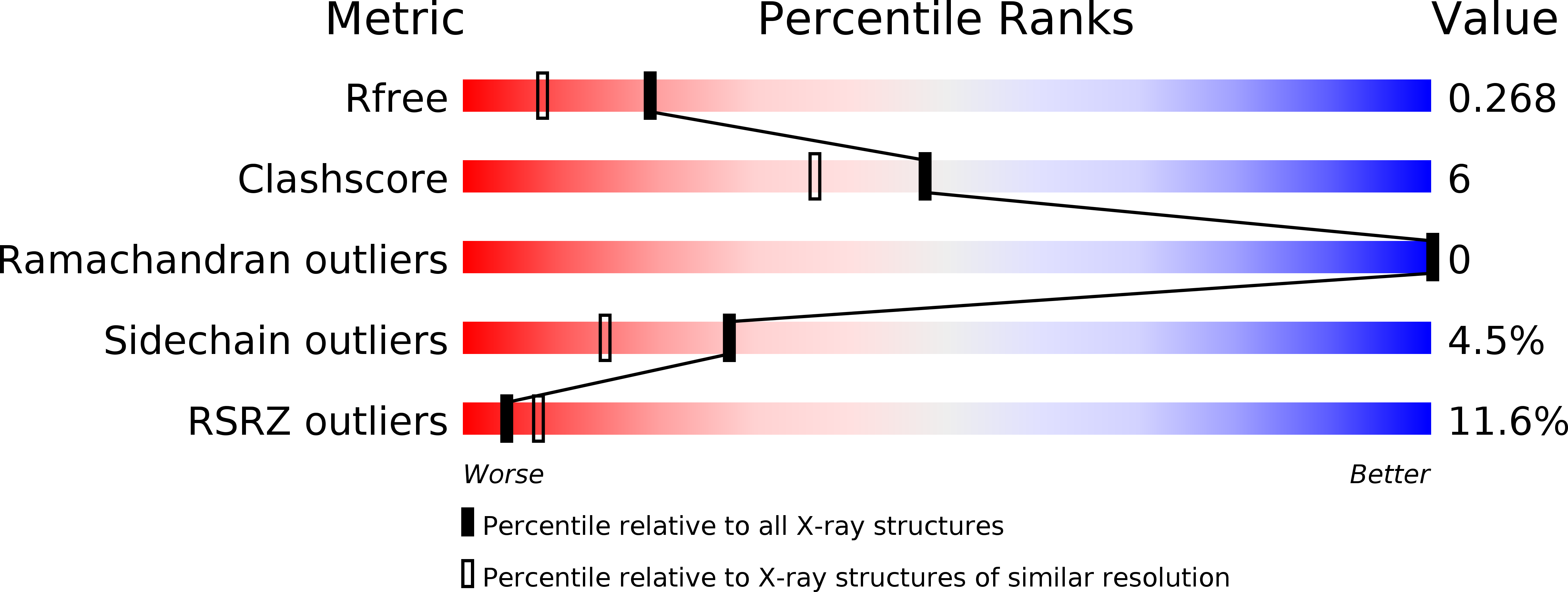
Resolution:
1.95 Å
R-Value Free:
0.25
R-Value Work:
0.20
R-Value Observed:
0.21
Space Group:
P 21 21 21
Macromolecular Entities
Polymer Type:polypeptide(L)
Molecule:Calmodulin-domain protein kinase 1
Gene (Uniprot):CDPK1
Chain IDs:A
Chain Length:508
Number of Molecules:1
Biological Source:Toxoplasma gondii
Primary Citation
Structures of apicomplexan calcium-dependent protein kinases reveal mechanism of activation by calcium.
Nat.Struct.Mol.Biol. 17 596 601 (2010)
PMID: 20436473 DOI: 10.1038/nsmb.1795

Abstact

Calcium-dependent protein kinases (CDPKs) have pivotal roles in the calcium-signaling pathway in plants, ciliates and apicomplexan parasites and comprise a calmodulin-dependent kinase (CaMK)-like kinase domain regulated by a calcium-binding domain in the C terminus. To understand this intramolecular mechanism of activation, we solved the structures of the autoinhibited (apo) and activated (calcium-bound) conformations of CDPKs from the apicomplexan parasites Toxoplasma gondii and Cryptosporidium parvum. In the apo form, the C-terminal CDPK activation domain (CAD) resembles a calmodulin protein with an unexpected long helix in the N terminus that inhibits the kinase domain in the same manner as CaMKII. Calcium binding triggers the reorganization of the CAD into a highly intricate fold, leading to its relocation around the base of the kinase domain to a site remote from the substrate binding site. This large conformational change constitutes a distinct mechanism in calcium signal-transduction pathways.

Legend

Protein

Chemical

Disease

Primary Citation of related structures