Deposition Date
2009-06-16
Release Date
2010-03-02
Last Version Date
2024-05-29
Entry Detail
PDB ID:
3HVN
Keywords:
Title:
Crystal structure of cytotoxin protein suilysin from Streptococcus suis
Biological Source:
Source Organism(s):
Streptococcus suis (Taxon ID: 391295)
Expression System(s):
Method Details:
Experimental Method:
Resolution:
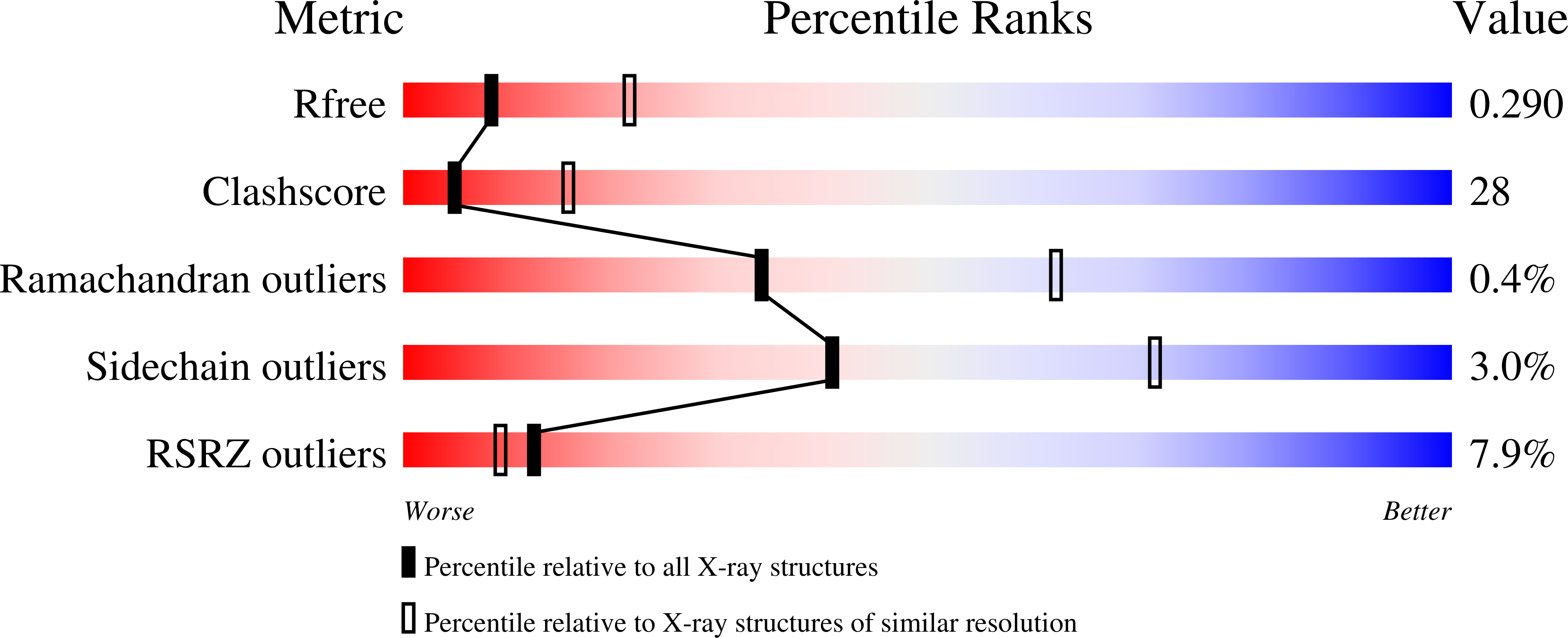
2.85 Å
R-Value Free:
0.29
R-Value Work:
0.27
R-Value Observed:
0.28
Space Group:
P 31 2 1