Deposition Date
2009-06-14
Release Date
2010-03-02
Last Version Date
2024-03-20
Entry Detail
PDB ID:
3HUG
Keywords:
Title:
Crystal structure of Mycobacterium tuberculosis anti-sigma factor RslA in complex with -35 promoter binding domain of sigL
Biological Source:
Source Organism(s):
Mycobacterium tuberculosis (Taxon ID: 83332)
Expression System(s):
Method Details:
Experimental Method:
Resolution:
2.35 Å
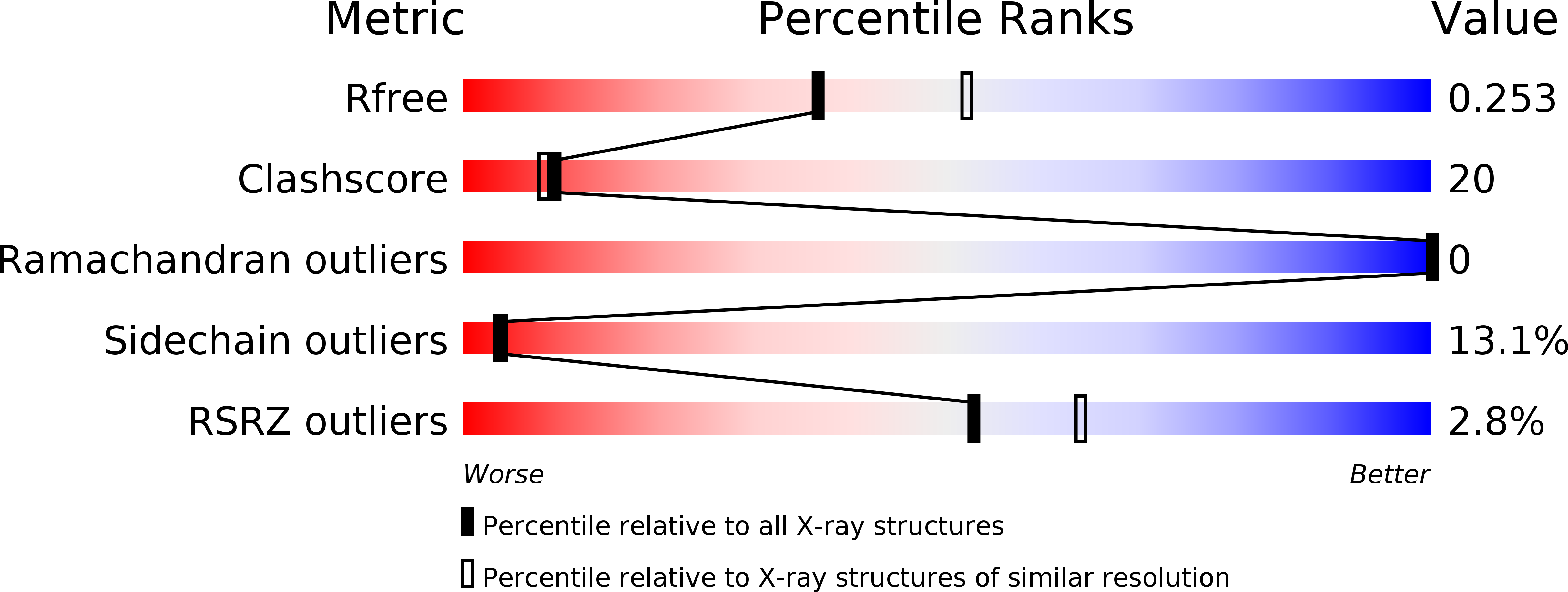
R-Value Free:
0.26
R-Value Work:
0.21
R-Value Observed:
0.21
Space Group:
P 21 21 21