3HTS image
Deposition Date 1998-11-16
Release Date 1999-04-29
Last Version Date 2023-09-06
Entry Detail
PDB ID:
3HTS
Title:
HEAT SHOCK TRANSCRIPTION FACTOR/DNA COMPLEX
Biological Source:
Source Organism:
Kluyveromyces lactis (Taxon ID: 28985)
(Taxon ID: )
Method Details:
Experimental Method:
Resolution:
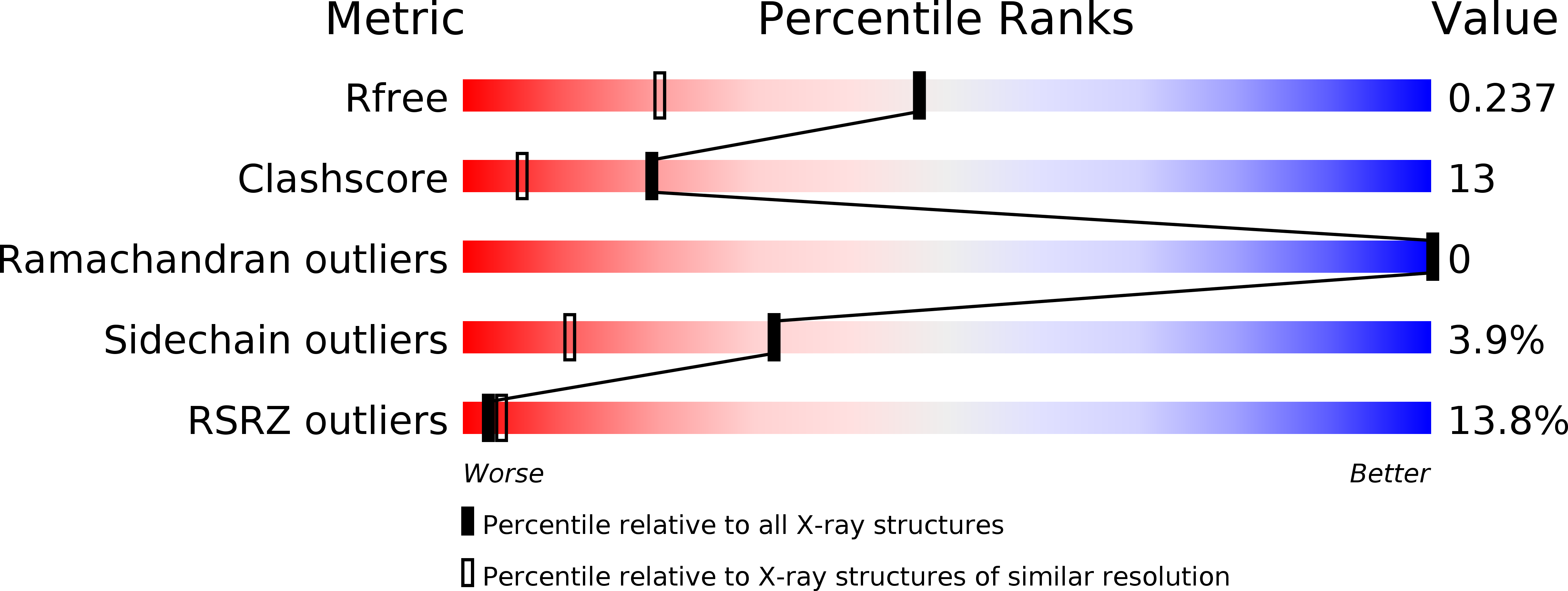
1.75 Å
R-Value Free:
0.24
R-Value Work:
0.20
Space Group:
C 1 2 1
Macromolecular Entities
Polymer Type:polydeoxyribonucleotide
Molecule:5'-D(*GP*GP*TP*TP*CP*TP*AP*GP*AP*AP*CP*C)-3'
Chain IDs:A
Chain Length:12
Number of Molecules:1
Biological Source:
Polymer Type:polypeptide(L)
Molecule:HEAT SHOCK TRANSCRIPTION FACTOR
Gene (Uniprot):HSF
Chain IDs:B
Chain Length:102
Number of Molecules:1
Biological Source:Kluyveromyces lactis
Ligand Molecules
Primary Citation
A new use for the 'wing' of the 'winged' helix-turn-helix motif in the HSF-DNA cocrystal.
Nat.Struct.Biol. 6 464 470 (1999)
PMID: 10331875 DOI: 10.1038/8269

Abstact

The 1.75 A crystal structure of the Kluyveromyces lactis heat shock transcription factor (HSF) DNA-binding domain (DBD) complexed with DNA reveals a protein-DNA interface with few direct major groove contacts and a number of phosphate backbone contacts that are primarily water-mediated interactions. The DBD, a 'winged' helix-turn-helix protein, displays a novel mode of binding in that the 'wing' does not contact DNA like all others of that class. Instead, the monomeric DBD, which crystallized as a symmetric dimer to a pair of nGAAn inverted repeats, uses the 'wing' to form part of the protein-protein contacts. This dimer interface is likely important for increasing the DNA-binding specificity and affinity of the trimeric form of HSF, as well as for increasing cooperativity between adjacent trimers.

Legend

Protein

Chemical

Disease

Primary Citation of related structures