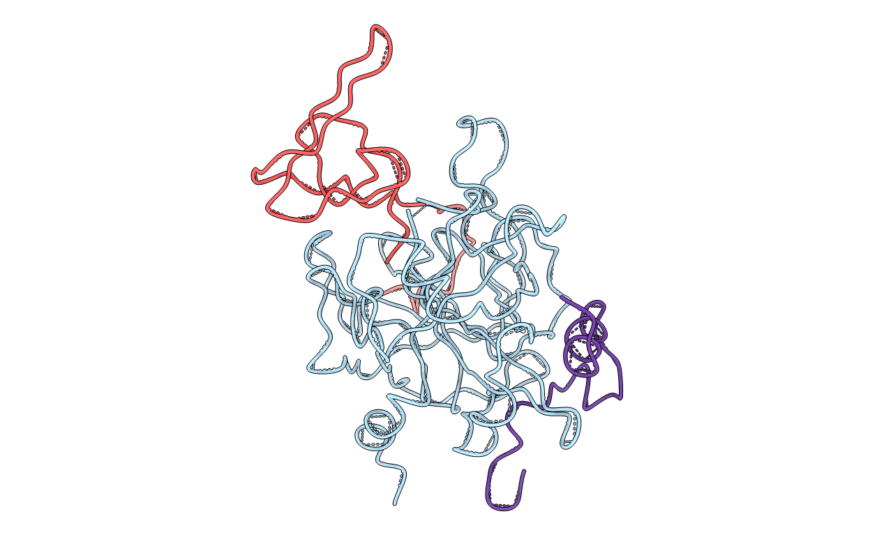
3HTC image
Entry Detail
PDB ID:
3HTC
Title:
THE STRUCTURE OF A COMPLEX OF RECOMBINANT HIRUDIN AND HUMAN ALPHA-THROMBIN
Biological Source:
PDB Version:
Deposition Date:
1993-06-11
Release Date:
1994-01-31
Method Details:
Experimental Method:
Resolution:
2.30 Å
R-Value Work:
0.19
Space Group:
P 43 21 2
Macromolecular Entities
Polymer Type:polypeptide(L)
Description:ALPHA-THROMBIN (LARGE SUBUNIT)
Chain IDs:B (auth: H)
Chain Length:259
Number of Molecules:1
Biological Source:Homo sapiens
Polymer Type:polypeptide(L)
Description:HIRUDIN VARIANT 2
Chain IDs:C (auth: I)
Chain Length:65
Number of Molecules:1
Biological Source:Hirudinaria manillensis
Polymer Type:polypeptide(L)
Description:ALPHA-THROMBIN (SMALL SUBUNIT)
Chain IDs:A (auth: L)
Chain Length:36
Number of Molecules:1
Biological Source:Homo sapiens
Ligand Molecules
Primary Citation
The structure of a complex of recombinant hirudin and human alpha-thrombin.
Science 249 277 280 (1990)
PMID: 2374926

Abstact

The crystallographic structure of a recombinant hirudin-thrombin complex has been solved at 2.3 angstrom (A) resolution. Hirudin consists of an NH2-terminal globular domain and a long (39 A) COOH-terminal extended domain. Residues Ile1 to Tyr3 of hirudin form a parallel beta-strand with Ser214 to Glu217 of thrombin with the nitrogen atom of Ile1 making a hydrogen bond with Ser195 O gamma atom of the catalytic site, but the specificity pocket of thrombin is not involved in the interaction. The COOH-terminal segment makes numerous electrostatic interactions with an anion-binding exosite of thrombin, whereas the last five residues are in a helical loop that forms many hydrophobic contacts. In all, 27 of the 65 residues of hirudin have contacts less than 4.0 A with thrombin (10 ion pairs and 23 hydrogen bonds). Such abundant interactions may account for the high affinity and specificity of hirudin.

Legend

Protein

Chemical

Disease

Primary Citation of related structures