Deposition Date
2009-06-11
Release Date
2009-10-13
Last Version Date
2024-03-20
Entry Detail
PDB ID:
3HT1
Keywords:
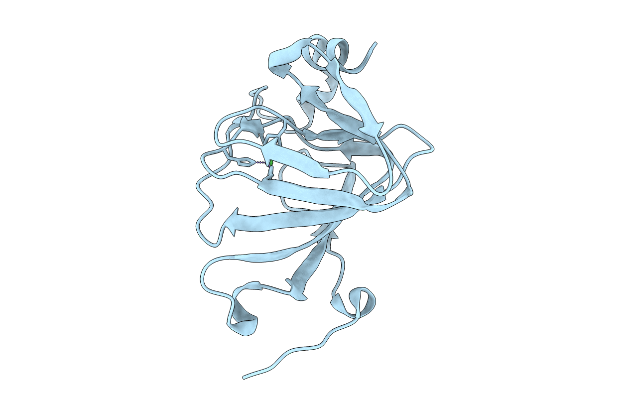
Title:
1.2A structure of the polyketide cyclase RemF from Streptomyces resistomycificus
Biological Source:
Source Organism(s):
Streptomyces resistomycificus (Taxon ID: 67356)
Expression System(s):
Method Details:
Experimental Method:
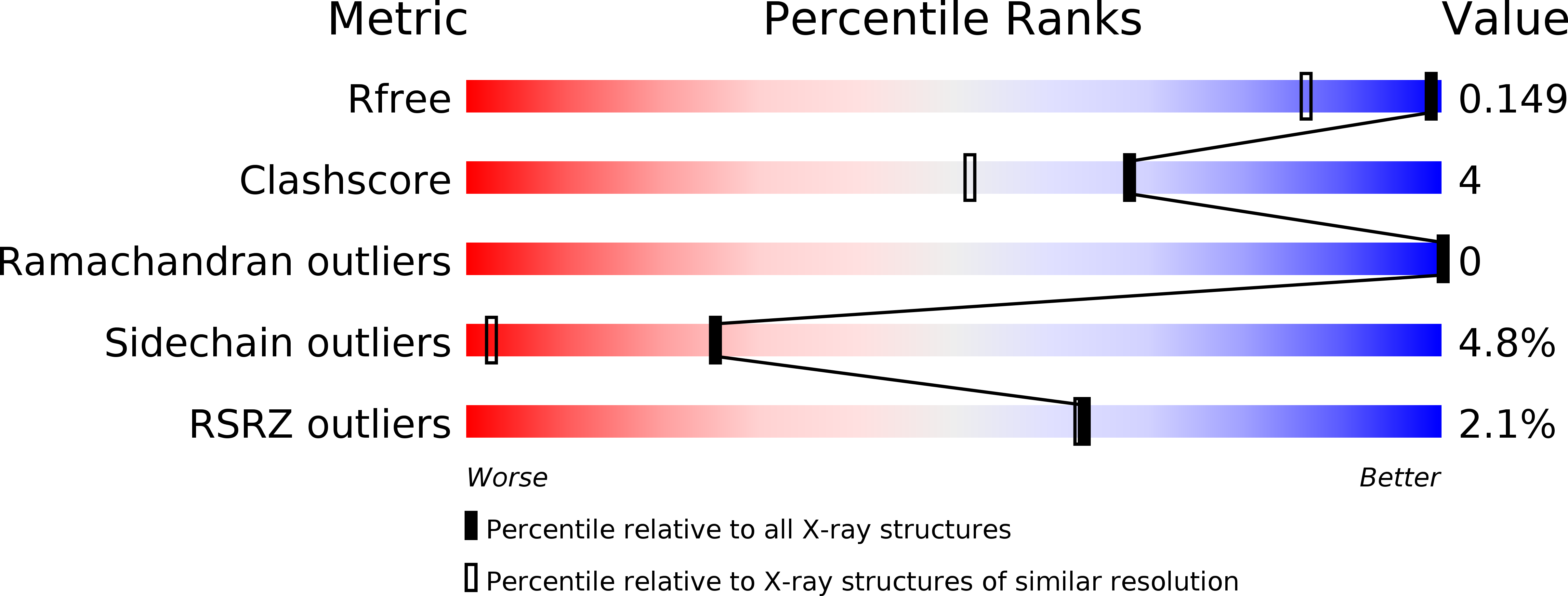
Resolution:
1.20 Å
R-Value Free:
0.17
R-Value Work:
0.12
R-Value Observed:
0.12
Space Group:
P 2 2 21