Deposition Date
2009-06-10
Release Date
2009-06-23
Last Version Date
2023-11-01
Entry Detail
PDB ID:
3HRW
Keywords:
Title:
Crystal structure of hemoglobin from mouse (Mus musculus)at 2.8
Biological Source:
Source Organism(s):
Mus musculus (Taxon ID: 10090)
Method Details:
Experimental Method:
Resolution:
2.80 Å
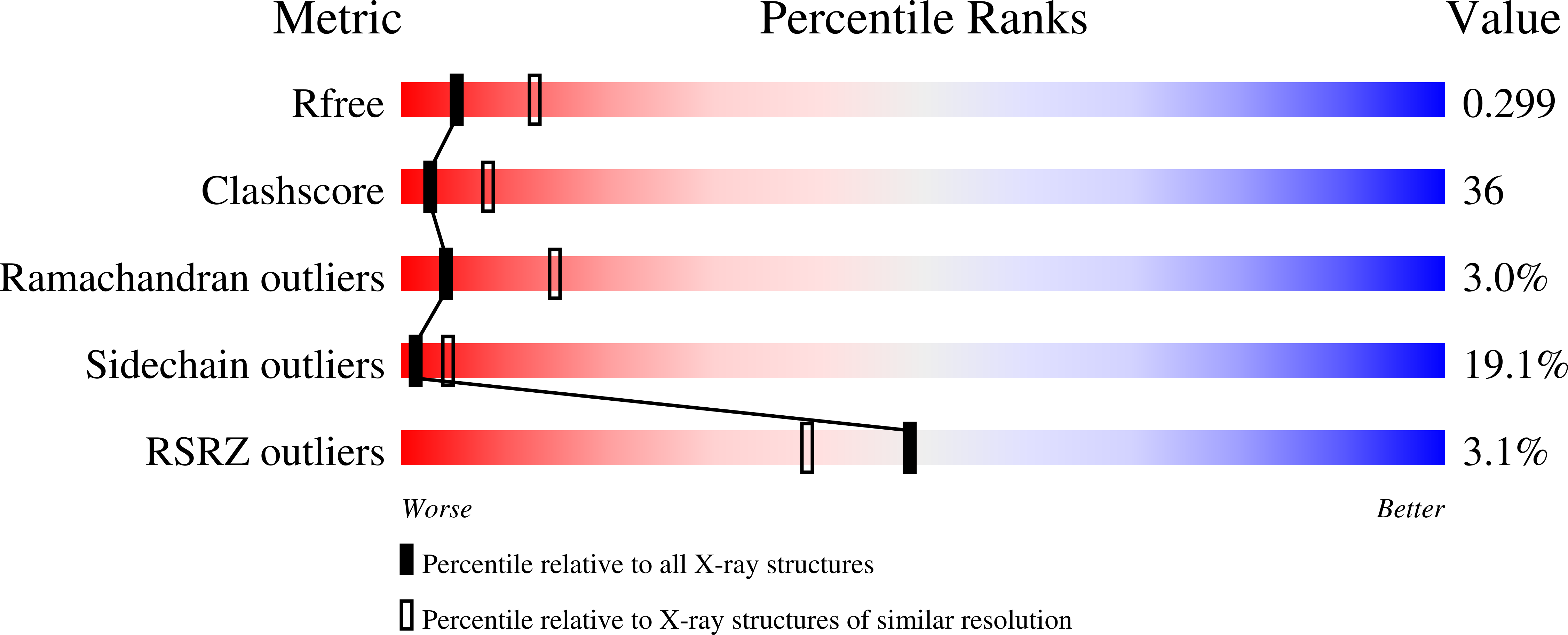
R-Value Free:
0.30
R-Value Work:
0.24
R-Value Observed:
0.24
Space Group:
P 21 21 21