3HOT image
Deposition Date 2009-06-03
Release Date 2009-09-22
Last Version Date 2024-11-06
Entry Detail
PDB ID:
3HOT
Title:
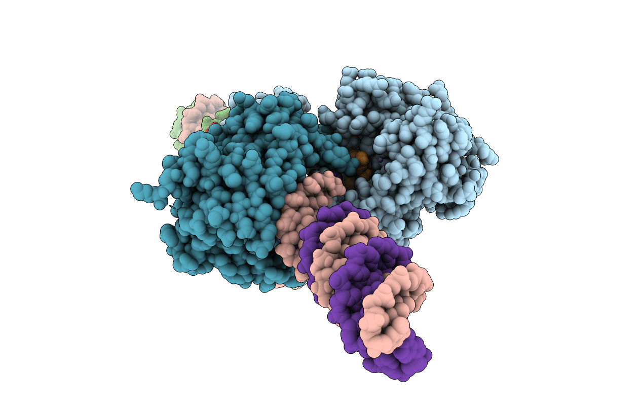
Crystal structure of the Mos1 mariner paired end complex with Mn
Biological Source:
Source Organism:
Host Organism:
Method Details:
Experimental Method:
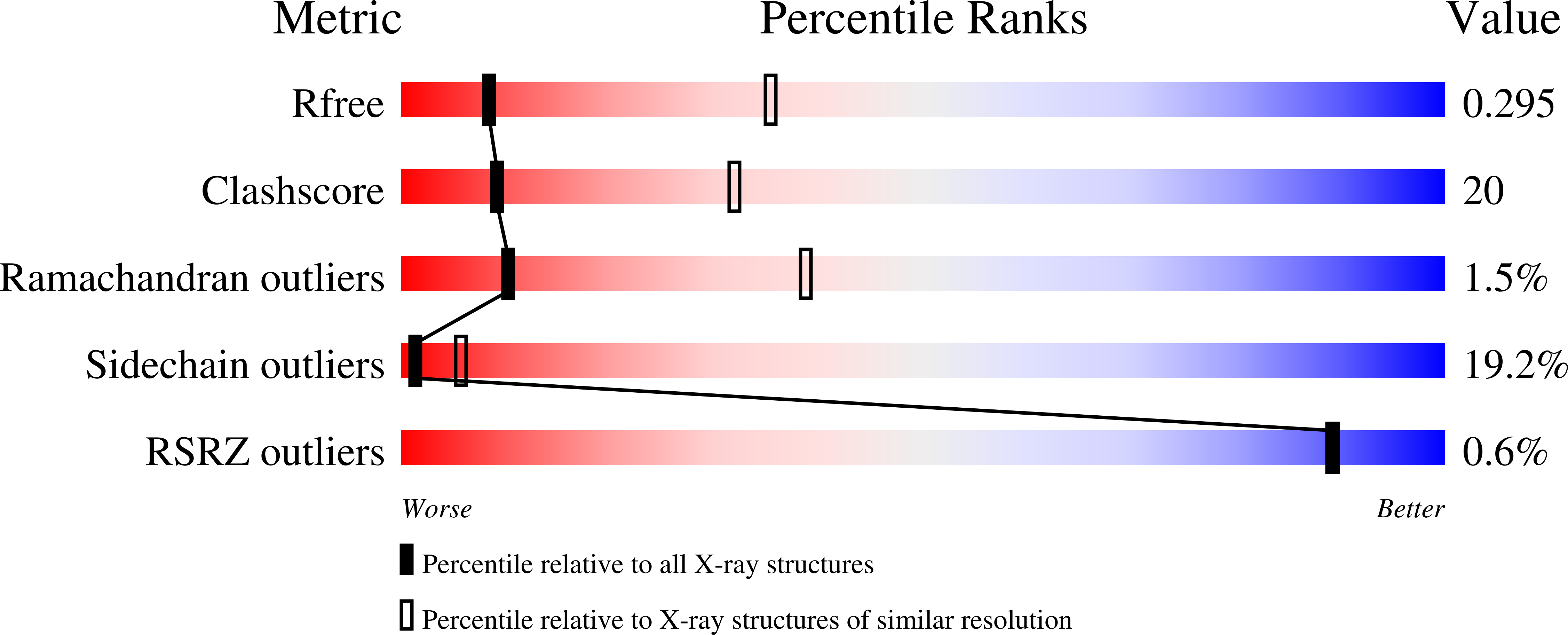
Resolution:
3.25 Å
R-Value Free:
0.30
R-Value Work:
0.24
R-Value Observed:
0.25
Space Group:
P 1 21 1
Macromolecular Entities
Structures with similar UniProt ID
Protein Blast
Polymer Type:polypeptide(L)
Molecule:Transposable element mariner, complete cds
Gene (Uniprot):mariner\T
Mutagens:T216A
Chain IDs:A, B
Chain Length:345
Number of Molecules:2
Biological Source:Drosophila mauritiana
Polymer Type:polydeoxyribonucleotide
Molecule:Mos1 NTS inverted repeat DNA
Chain IDs:C, E, G
Chain Length:25
Number of Molecules:3
Biological Source:synthetic construct
Polymer Type:polydeoxyribonucleotide
Molecule:Mos1 TS inverted repeat DNA
Chain IDs:D, F, H
Chain Length:28
Number of Molecules:3
Biological Source:synthetic construct
Modified Residue
Compound ID Chain ID Parent Comp ID Details 2D Image
5IU C DU 5-IODO-2'-DEOXYURIDINE-5'-MONOPHOSPHATE
Primary Citation
Molecular architecture of the Mos1 paired-end complex: the structural basis of DNA transposition in a eukaryote
Cell(Cambridge,Mass.) 138 1096 1108 (2009)
PMID: 19766564 DOI: 10.1016/j.cell.2009.07.012

Abstact

A key step in cut-and-paste DNA transposition is the pairing of transposon ends before the element is excised and inserted at a new site in its host genome. Crystallographic analyses of the paired-end complex (PEC) formed from precleaved transposon ends and the transposase of the eukaryotic element Mos1 reveals two parallel ends bound to a dimeric enzyme. The complex has a trans arrangement, with each transposon end recognized by the DNA binding region of one transposase monomer and by the active site of the other monomer. Two additional DNA duplexes in the crystal indicate likely binding sites for flanking DNA. Biochemical data provide support for a model of the target capture complex and identify Arg186 to be critical for target binding. Mixing experiments indicate that a transposase dimer initiates first-strand cleavage and suggest a pathway for PEC formation.

Legend

Protein

Chemical

Disease

Primary Citation of related structures
Feedback Form
Name
Email
Institute
Feedback