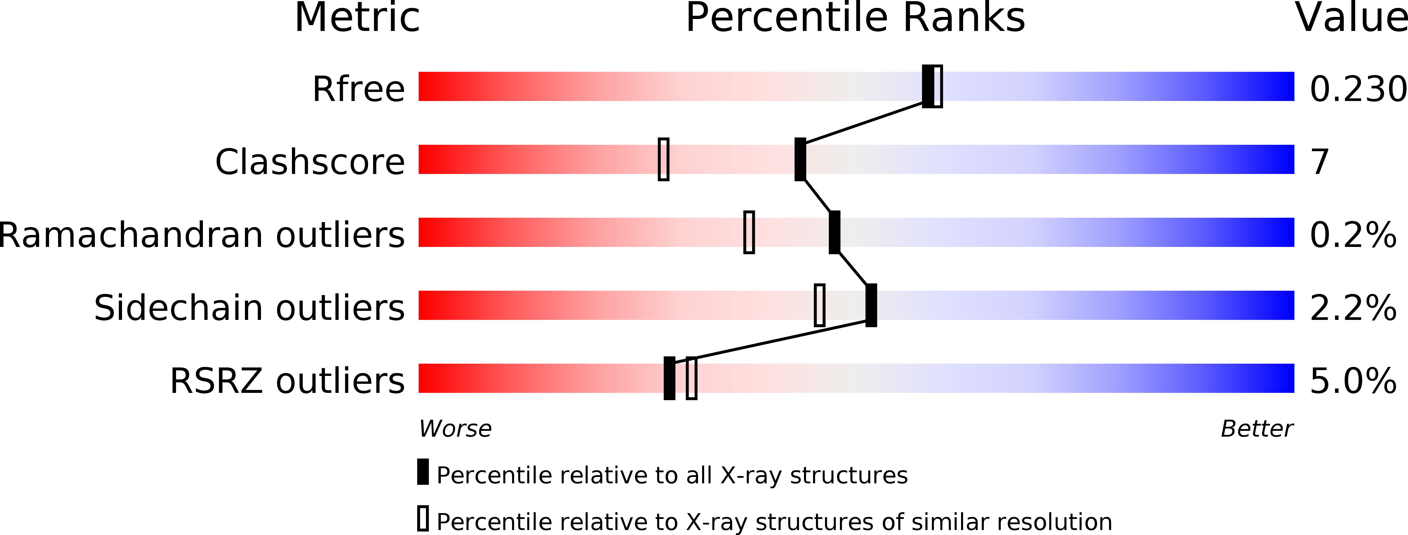
3HOL image
Deposition Date 2009-06-02
Release Date 2009-10-06
Last Version Date 2024-10-16
Entry Detail
PDB ID:
3HOL
Title:
The Structure of Intact Ap-TbpB (N and C lobes)
Biological Source:
Source Organism:
Host Organism:
Method Details:
Experimental Method:
Resolution:
1.98 Å
R-Value Free:
0.23
R-Value Work:
0.18
R-Value Observed:
0.18
Space Group:
P 21 21 21
Macromolecular Entities
Polymer Type:polypeptide(L)
Molecule:TbpB
Gene (Uniprot):tbpB
Chain IDs:A
Chain Length:509
Number of Molecules:1
Biological Source:Actinobacillus pleuropneumoniae
Primary Citation
Insights into the bacterial transferrin receptor: the structure of transferrin-binding protein B from Actinobacillus pleuropneumoniae.
Mol.Cell 35 523 533 (2009)
PMID: 19716795 DOI: 10.1016/j.molcel.2009.06.029

Abstact

Pathogenic bacteria from the Neisseriaceae and Pasteurellacea families acquire iron directly from the host iron-binding glycoprotein, transferrin (Tf), in a process mediated by surface receptor proteins that directly bind host Tf, extract the iron, and transport it across the outer membrane. The bacterial Tf receptor is comprised of a surface exposed lipoprotein, Tf-binding protein B (TbpB), and an integral outer-membrane protein, Tf-binding protein A (TbpA), both of which are essential for survival in the host. In this study, we report the 1.98 A resolution structure of TbpB from the porcine pathogen Actinobacillus pleuropneumoniae, providing insights into the mechanism of Tf binding and the role of TbpB. A model for the complex of TbpB bound to Tf is proposed. Mutation of a single surface-exposed Phe residue on TbpB within the predicted interface completely abolishes binding to Tf, suggesting that the TbpB N lobe comprises the sole high-affinity binding region for Tf.

Legend

Protein

Chemical

Disease

Primary Citation of related structures