3HO8 image
Deposition Date 2009-06-01
Release Date 2009-06-30
Last Version Date 2024-02-21
Entry Detail
PDB ID:
3HO8
Keywords:
Title:
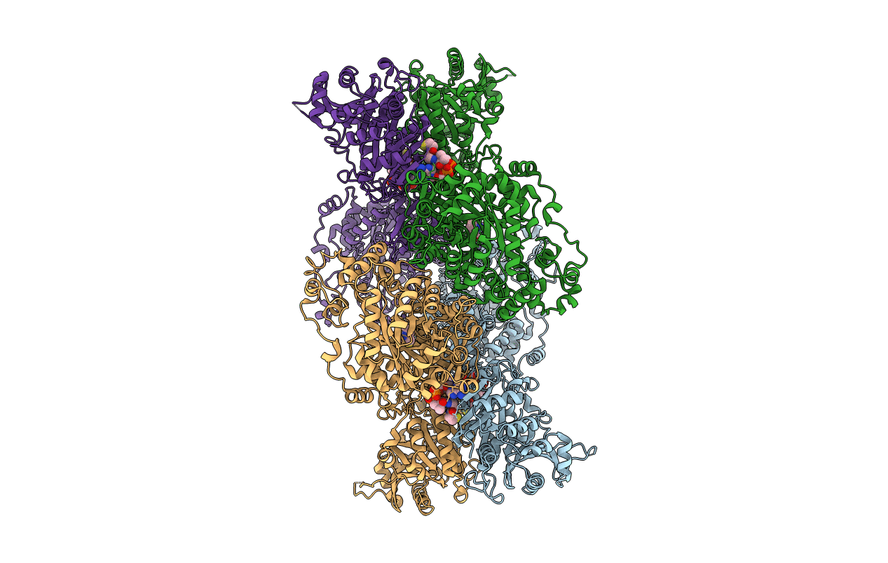
Crystal Structure of S. aureus Pyruvate Carboxylase in complex with Coenzyme A
Biological Source:
Host Organism:
Method Details:
Experimental Method:
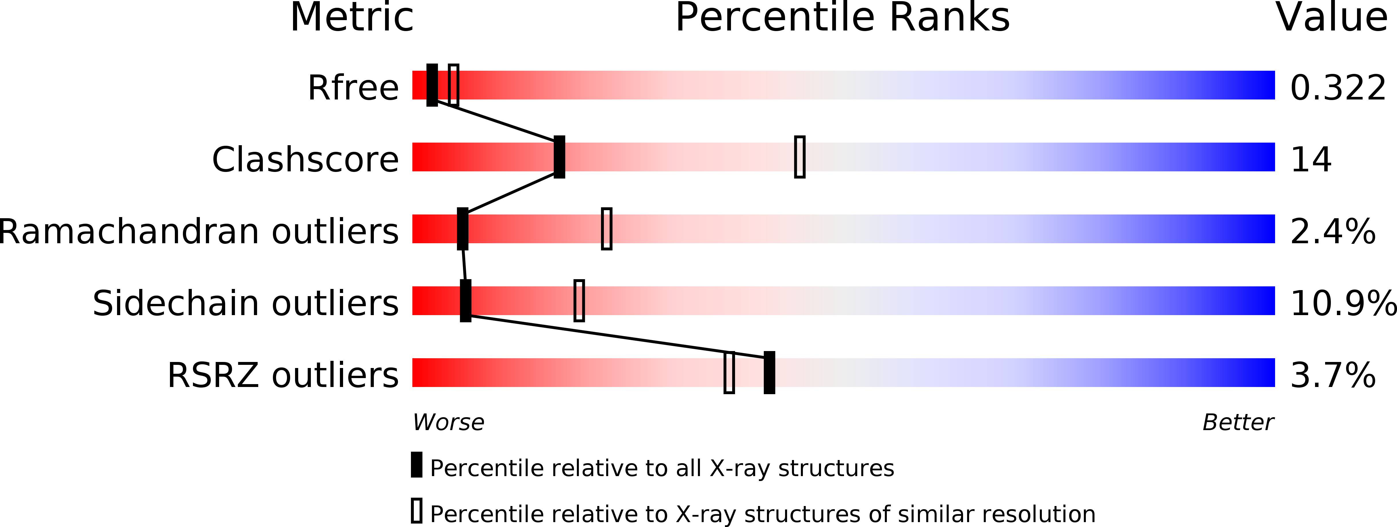
Resolution:
2.90 Å
R-Value Free:
0.32
R-Value Work:
0.26
R-Value Observed:
0.26
Space Group:
P 21 21 21
Macromolecular Entities
Structures with similar UniProt ID
Protein Blast
Polymer Type:polypeptide(L)
Molecule:Pyruvate carboxylase
Chain IDs:A, B (auth: D), C, D (auth: B)
Chain Length:1150
Number of Molecules:4
Biological Source:Staphylococcus aureus subsp. aureus Mu50
Primary Citation
A Symmetrical Tetramer for S. aureus Pyruvate Carboxylase in Complex with Coenzyme A.
Structure 17 823 832 (2009)
PMID: 19523900 DOI: 10.1016/j.str.2009.04.008

Abstact

Pyruvate carboxylase (PC) is a conserved metabolic enzyme with important cellular functions. We report crystallographic and cryo-electron microscopy (EM) studies of Staphylococcus aureus PC (SaPC) in complex with acetyl-CoA, an allosteric activator, and mutagenesis, biochemical, and structural studies of the biotin binding site of its carboxyltransferase (CT) domain. The disease-causing A610T mutation abolishes catalytic activity by blocking biotin binding to the CT active site, and Thr908 might play a catalytic role in the CT reaction. The crystal structure of SaPC in complex with CoA reveals a symmetrical tetramer, with one CoA molecule bound to each monomer, and cryo-EM studies confirm the symmetrical nature of the tetramer. These observations are in sharp contrast to the highly asymmetrical tetramer of Rhizobium etli PC in complex with ethyl-CoA. Our structural information suggests that acetyl-CoA promotes a conformation for the dimer of the biotin carboxylase domain of PC that might be catalytically more competent.

Legend

Protein

Chemical

Disease

Primary Citation of related structures
Feedback Form
Name
Email
Institute
Feedback