
Deposition Date
2009-05-29
Release Date
2009-06-16
Last Version Date
2024-10-30
Entry Detail
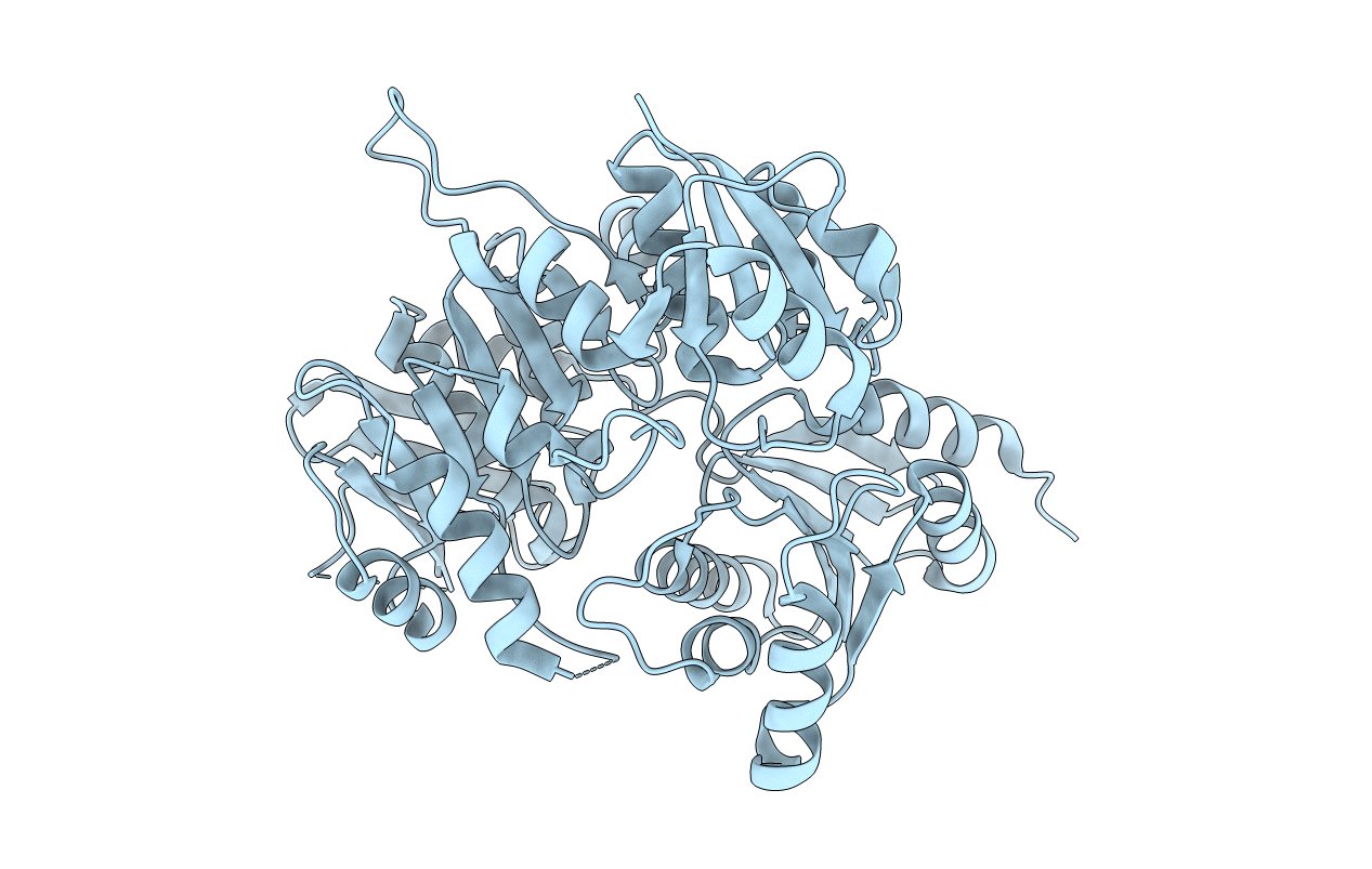
PDB ID:
3HN7
Keywords:
Title:
Crystal structure of a murein peptide ligase mpl (psyc_0032) from psychrobacter arcticus 273-4 at 1.65 A resolution
Biological Source:
Source Organism(s):
Psychrobacter arcticus 273-4 (Taxon ID: 259536)
Expression System(s):
Method Details:
Experimental Method:
Resolution:
1.65 Å
R-Value Free:
0.18
R-Value Work:
0.15
R-Value Observed:
0.15
Space Group:
C 1 2 1
