3HLJ image
Deposition Date 2009-05-27
Release Date 2010-03-02
Last Version Date 2023-11-01
Entry Detail
PDB ID:
3HLJ
Keywords:
Title:
Crystal structure of human carbonic anhydrase isozyme II with 3-methylthiobenzimidazo[1,2-c][1,2,3]thiadiazol-7-sulfonamide
Biological Source:
Source Organism:
Homo sapiens (Taxon ID: 9606)
Host Organism:
Method Details:
Experimental Method:
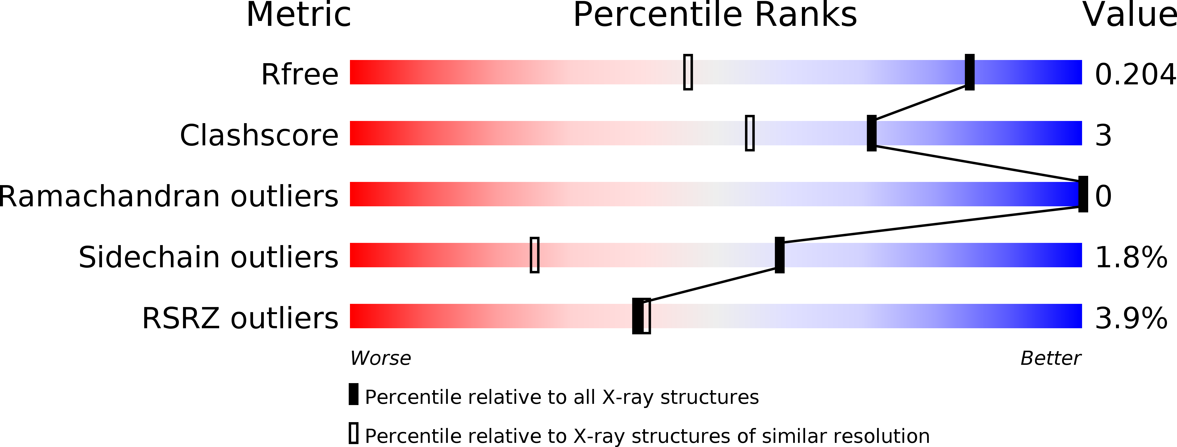
Resolution:
1.44 Å
R-Value Free:
0.20
R-Value Work:
0.17
R-Value Observed:
0.17
Space Group:
P 1 21 1
Macromolecular Entities
Structures with similar UniProt ID
Protein Blast
Polymer Type:polypeptide(L)
Molecule:Carbonic anhydrase 2
Gene (Uniprot):CA2
Chain IDs:A
Chain Length:260
Number of Molecules:1
Biological Source:Homo sapiens
Primary Citation
Inhibition and binding studies of carbonic anhydrase isozymes I, II and IX with benzimidazo[1,2-c][1,2,3]thiadiazole-7-sulphonamides
J Enzyme Inhib Med Chem 25 863 870 (2010)
PMID: 20166809 DOI: 10.3109/14756360903571685

Abstact

The binding and inhibition strength of a series of benzimidazo[1,2-c][1,2,3]thiadiazole-7-sulphonamides were determined for recombinant human carbonic anhydrase isoforms I, II, and IX. The inhibition strength was determined by a stop-flow method to measure carbon dioxide hydration. Inhibitor-enzyme binding was determined by two biophysical techniques--isothermal titration calorimetry and thermal shift assay. The co-crystal structure was determined by X-ray crystallography. Comparing the results obtained using three different inhibition and binding methods increased the accuracy of compound affinity ranking and the ability to determine compound inhibitory specificity towards a particular carbonic anhydrase isoform. In most cases, all three methods yielded the same results despite using very different approaches to measure the binding and inhibition reactions. Some of the compounds studied are submicromolar inhibitors of the isoform IX, a prominent cancer target.

Legend

Protein

Chemical

Disease

Primary Citation of related structures