3HKL image
Deposition Date 2009-05-24
Release Date 2009-08-25
Last Version Date 2024-10-30
Entry Detail
PDB ID:
3HKL
Title:
Crystal Structure of the Frizzled-like Cysteine-rich Domain of MuSK
Biological Source:
Source Organism:
Host Organism:
Method Details:
Experimental Method:
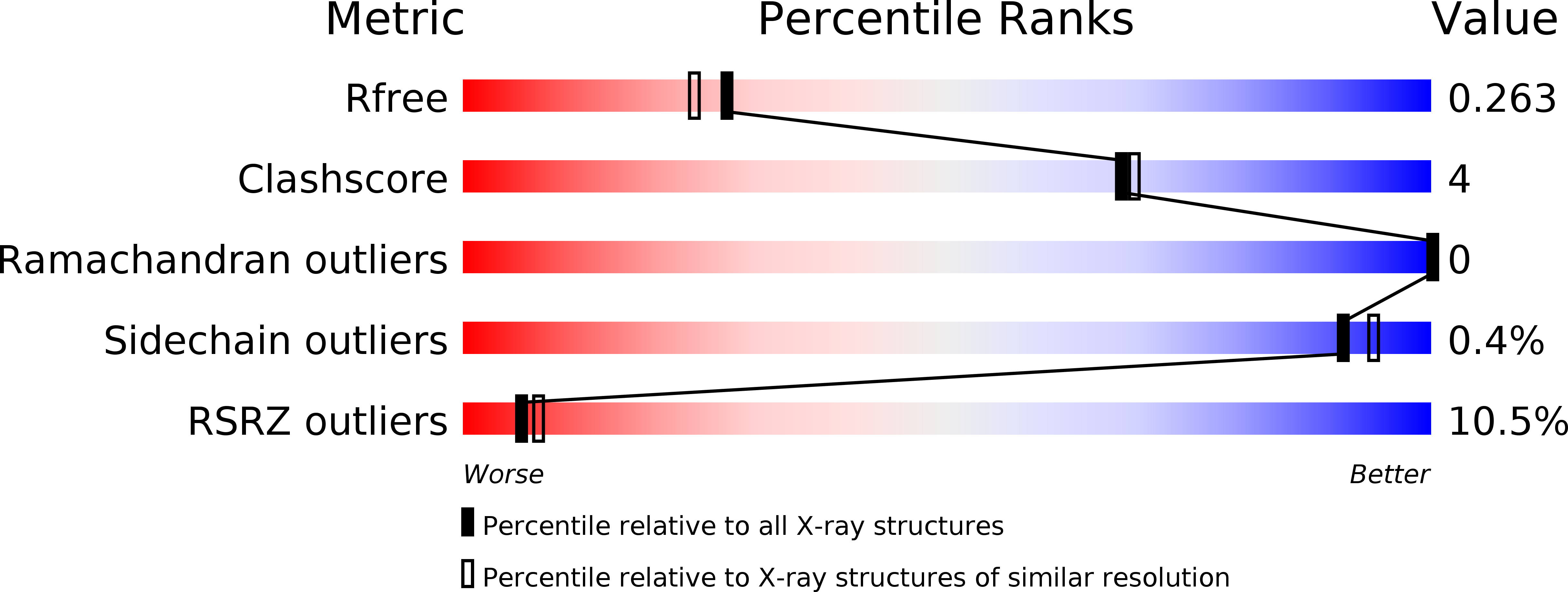
Resolution:
2.10 Å
R-Value Free:
0.26
R-Value Work:
0.22
R-Value Observed:
0.22
Space Group:
P 1 21 1
Macromolecular Entities
Structures with similar UniProt ID
Protein Blast
Polymer Type:polypeptide(L)
Molecule:Muscle, skeletal receptor tyrosine protein kinase
Gene (Uniprot):Musk
Chain IDs:A, B
Chain Length:197
Number of Molecules:2
Biological Source:Rattus norvegicus
Modified Residue
Compound ID Chain ID Parent Comp ID Details 2D Image
ASN A ASN GLYCOSYLATION SITE
Primary Citation
Crystal structure of the frizzled-like cysteine-rich domain of the receptor tyrosine kinase MuSK.
J.Mol.Biol. 393 1 9 (2009)
PMID: 19664639 DOI: 10.1016/j.jmb.2009.07.091

Abstact

Muscle-specific kinase (MuSK) is an essential receptor tyrosine kinase for the establishment and maintenance of the neuromuscular junction (NMJ). Activation of MuSK by agrin, a neuronally derived heparan-sulfate proteoglycan, and LRP4 (low-density lipoprotein receptor-related protein-4), the agrin receptor, leads to clustering of acetylcholine receptors on the postsynaptic side of the NMJ. The ectodomain of MuSK comprises three immunoglobulin-like domains and a cysteine-rich domain (Fz-CRD) related to those in Frizzled proteins, the receptors for Wnts. Here, we report the crystal structure of the MuSK Fz-CRD at 2.1 A resolution. The structure reveals a five-disulfide-bridged domain similar to CRDs of Frizzled proteins but with a divergent C-terminal region. An asymmetric dimer present in the crystal structure implicates surface hydrophobic residues that may function in homotypic or heterotypic interactions to mediate co-clustering of MuSK, rapsyn, and acetylcholine receptors at the NMJ.

Legend

Protein

Chemical

Disease

Primary Citation of related structures