3HK3 image
Deposition Date 2009-05-22
Release Date 2009-07-07
Last Version Date 2024-10-30
Entry Detail
PDB ID:
3HK3
Keywords:
Title:
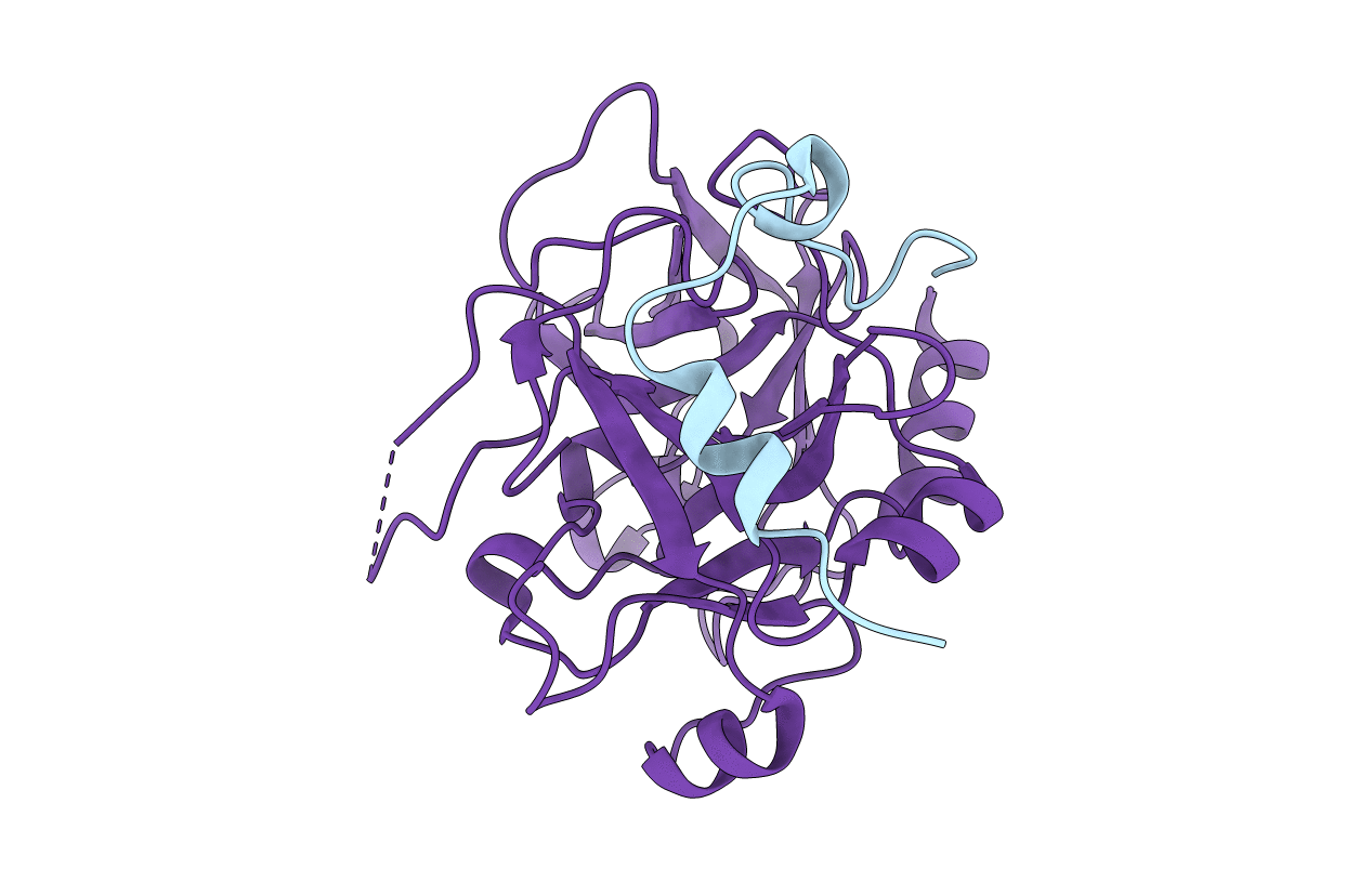
Crystal structure of murine thrombin mutant W215A/E217A (one molecule in the asymmetric unit)
Biological Source:
Source Organism:
Mus musculus (Taxon ID: 10090)
Host Organism:
Method Details:
Experimental Method:
Resolution:
1.94 Å
R-Value Free:
0.23
R-Value Work:
0.18
R-Value Observed:
0.18
Space Group:
P 21 21 21
Macromolecular Entities
Structures with similar UniProt ID
Protein Blast
Polymer Type:polypeptide(L)
Molecule:Thrombin light chain
Gene (Uniprot):F2
Chain IDs:A
Chain Length:44
Number of Molecules:1
Biological Source:Mus musculus
Structures with similar UniProt ID
Protein Blast
Polymer Type:polypeptide(L)
Molecule:Thrombin heavy chain
Gene (Uniprot):F2
Mutagens:W215A, E217A
Chain IDs:B
Chain Length:258
Number of Molecules:1
Biological Source:Mus musculus
Primary Citation
Mechanism of the Anticoagulant Activity of Thrombin Mutant W215A/E217A.
J.Biol.Chem. 284 24098 24105 (2009)
PMID: 19586901 DOI: 10.1074/jbc.M109.025403

Abstact

The thrombin mutant W215A/E217A (WE) is a potent anticoagulant both in vitro and in vivo. Previous x-ray structural studies have shown that WE assumes a partially collapsed conformation that is similar to the inactive E* form, which explains its drastically reduced activity toward substrate. Whether this collapsed conformation is genuine, rather than the result of crystal packing or the mutation introduced in the critical 215-217 beta-strand, and whether binding of thrombomodulin to exosite I can allosterically shift the E* form to the active E form to restore activity toward protein C are issues of considerable mechanistic importance to improve the design of an anticoagulant thrombin mutant for therapeutic applications. Here we present four crystal structures of WE in the human and murine forms that confirm the collapsed conformation reported previously under different experimental conditions and crystal packing. We also present structures of human and murine WE bound to exosite I with a fragment of the platelet receptor PAR1, which is unable to shift WE to the E form. These structural findings, along with kinetic and calorimetry data, indicate that WE is strongly stabilized in the E* form and explain why binding of ligands to exosite I has only a modest effect on the E*-E equilibrium for this mutant. The E* --> E transition requires the combined binding of thrombomodulin and protein C and restores activity of the mutant WE in the anticoagulant pathway.

Legend

Protein

Chemical

Disease

Primary Citation of related structures