3HJK image
Deposition Date 2009-05-21
Release Date 2009-09-29
Last Version Date 2023-09-06
Entry Detail
PDB ID:
3HJK
Title:
2.0 Angstrom Structure of the Ile74Val Variant of Vivid (VVD).
Biological Source:
Source Organism:
Host Organism:
Method Details:
Experimental Method:
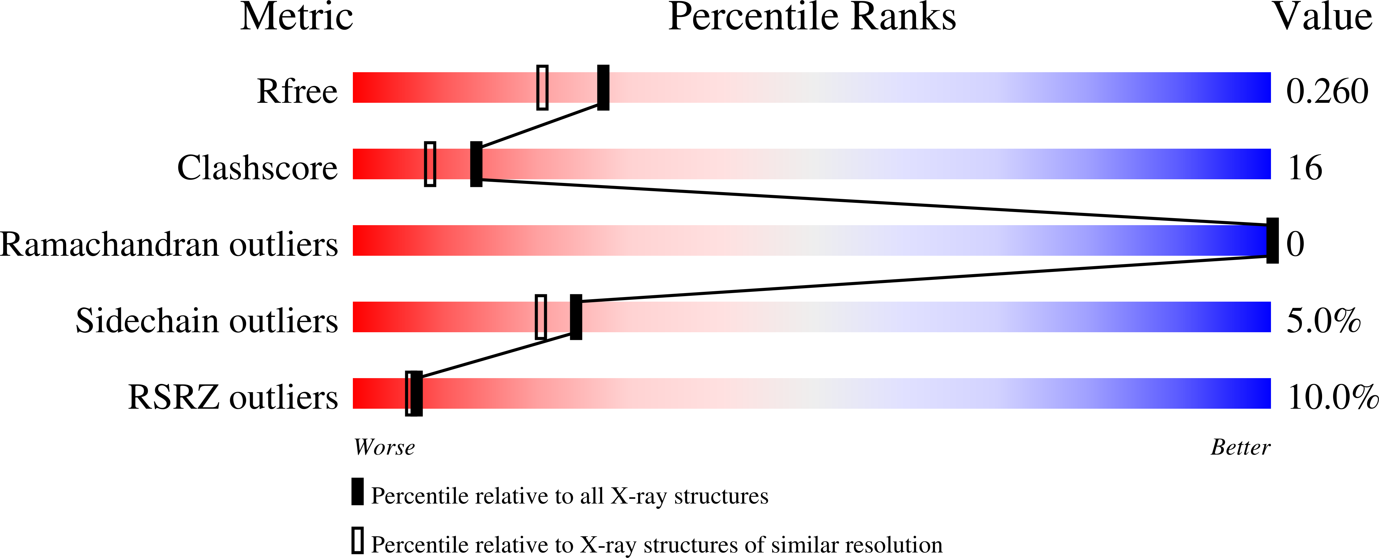
Resolution:
2.00 Å
R-Value Free:
0.26
R-Value Work:
0.24
R-Value Observed:
0.26
Space Group:
P 1 21 1
Macromolecular Entities
Polymer Type:polypeptide(L)
Molecule:Vivid PAS protein VVD
Gene (Uniprot):vvd
Mutations:I74V
Chain IDs:A, B
Chain Length:154
Number of Molecules:2
Biological Source:Neurospora crassa
Ligand Molecules
Primary Citation
Mechanism-based tuning of a LOV domain photoreceptor.
Nat.Chem.Biol. 5 827 834 (2009)
PMID: 19718042 DOI: 10.1038/nchembio.210

Abstact

Phototropin-like LOV domains form a cysteinyl-flavin adduct in response to blue light but show considerable variation in output signal and the lifetime of the photo-adduct signaling state. Mechanistic studies of the slow-cycling fungal LOV photoreceptor Vivid (VVD) reveal the importance of reactive cysteine conformation, flavin electronic environment and solvent accessibility for adduct scission and thermal reversion. Proton inventory, pH effects, base catalysis and structural studies implicate flavin N(5) deprotonation as rate-determining for recovery. Substitutions of active site residues Ile74, Ile85, Met135 and Met165 alter photoadduct lifetimes by over four orders of magnitude in VVD, and similar changes in other LOV proteins show analogous effects. Adduct state decay rates also correlate with changes in conformational and oligomeric properties of the protein necessary for signaling. These findings link natural sequence variation of LOV domains to function and provide a means to design broadly reactive light-sensitive probes.

Legend

Protein

Chemical

Disease

Primary Citation of related structures