3HHI image
Deposition Date 2009-05-15
Release Date 2010-05-26
Last Version Date 2024-11-20
Entry Detail
PDB ID:
3HHI
Keywords:
Title:
Crystal Structure of Cathepsin B from T. brucei in complex with CA074
Biological Source:
Source Organism:
Host Organism:
Method Details:
Experimental Method:
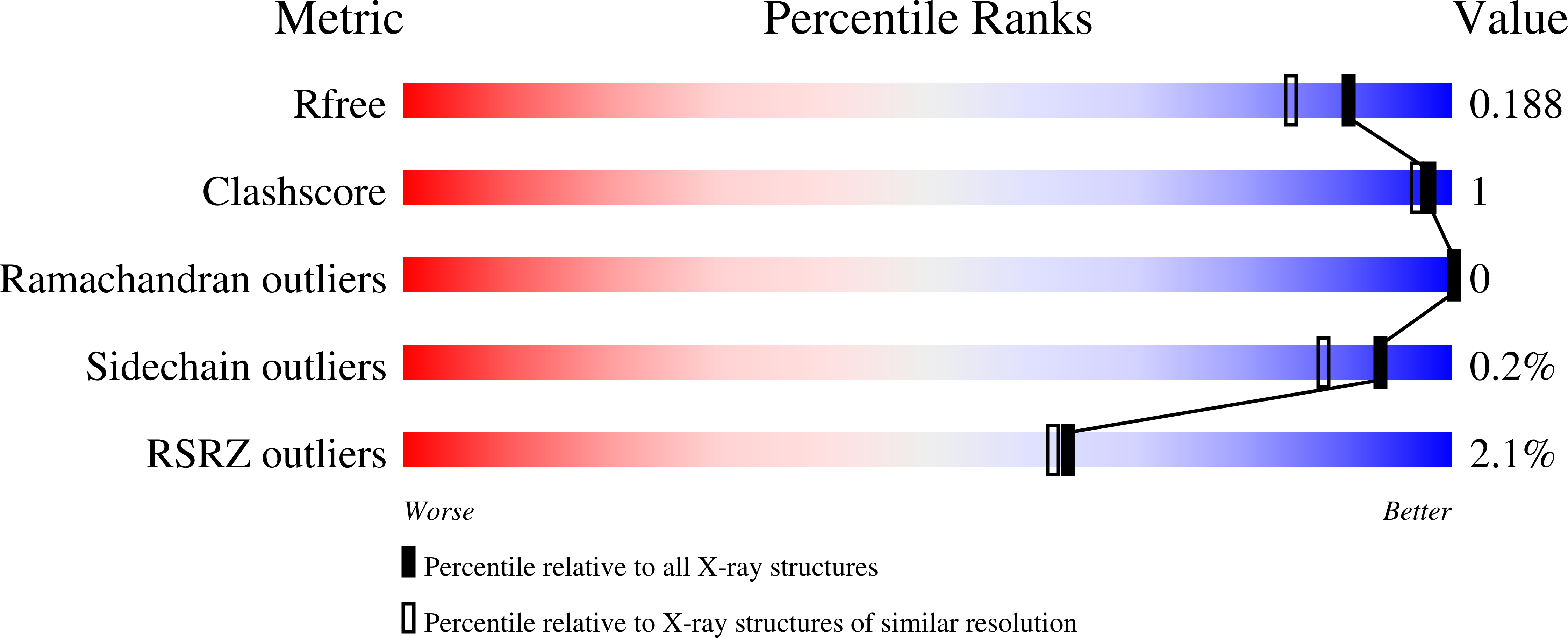
Resolution:
1.60 Å
R-Value Free:
0.17
R-Value Work:
0.14
R-Value Observed:
0.14
Space Group:
P 1 21 1
Macromolecular Entities
Structures with similar UniProt ID
Protein Blast
Polymer Type:polypeptide(L)
Molecule:Cathepsin B-like cysteine protease
Mutagens:N216D
Chain IDs:A, B
Chain Length:325
Number of Molecules:2
Biological Source:Trypanosoma brucei
Peptide-like Molecules
PRD_001032
Primary Citation
Crystal Structures of TbCatB and rhodesain, potential chemotherapeutic targets and major cysteine proteases of Trypanosoma brucei
Plos Negl Trop Dis 4 e701 e701 (2010)
PMID: 20544024 DOI: 10.1371/journal.pntd.0000701

Abstact

BACKGROUND Trypanosoma brucei is the etiological agent of Human African Trypanosomiasis, an endemic parasitic disease of sub-Saharan Africa. TbCatB and rhodesain are the sole Clan CA papain-like cysteine proteases produced by the parasite during infection of the mammalian host and are implicated in the progression of disease. Of considerable interest is the exploration of these two enzymes as targets for cysteine protease inhibitors that are effective against T. brucei. METHODS AND FINDINGS We have determined, by X-ray crystallography, the first reported structure of TbCatB in complex with the cathepsin B selective inhibitor CA074. In addition we report the structure of rhodesain in complex with the vinyl-sulfone K11002. CONCLUSIONS The mature domain of our TbCat*CA074 structure contains unique features for a cathepsin B-like enzyme including an elongated N-terminus extending 16 residues past the predicted maturation cleavage site. N-terminal Edman sequencing reveals an even longer extension than is observed amongst the ordered portions of the crystal structure. The TbCat*CA074 structure confirms that the occluding loop, which is an essential part of the substrate-binding site, creates a larger prime side pocket in the active site cleft than is found in mammalian cathepsin B-small molecule structures. Our data further highlight enhanced flexibility in the occluding loop main chain and structural deviations from mammalian cathepsin B enzymes that may affect activity and inhibitor design. Comparisons with the rhodesain*K11002 structure highlight key differences that may impact the design of cysteine protease inhibitors as anti-trypanosomal drugs.

Legend

Protein

Chemical

Disease

Primary Citation of related structures