3HGV image
Deposition Date 2009-05-14
Release Date 2010-04-28
Last Version Date 2024-10-30
Entry Detail
PDB ID:
3HGV
Title:
Structure of Phenazine Antibiotic Biosynthesis Protein
Biological Source:
Source Organism:
Host Organism:
Method Details:
Experimental Method:
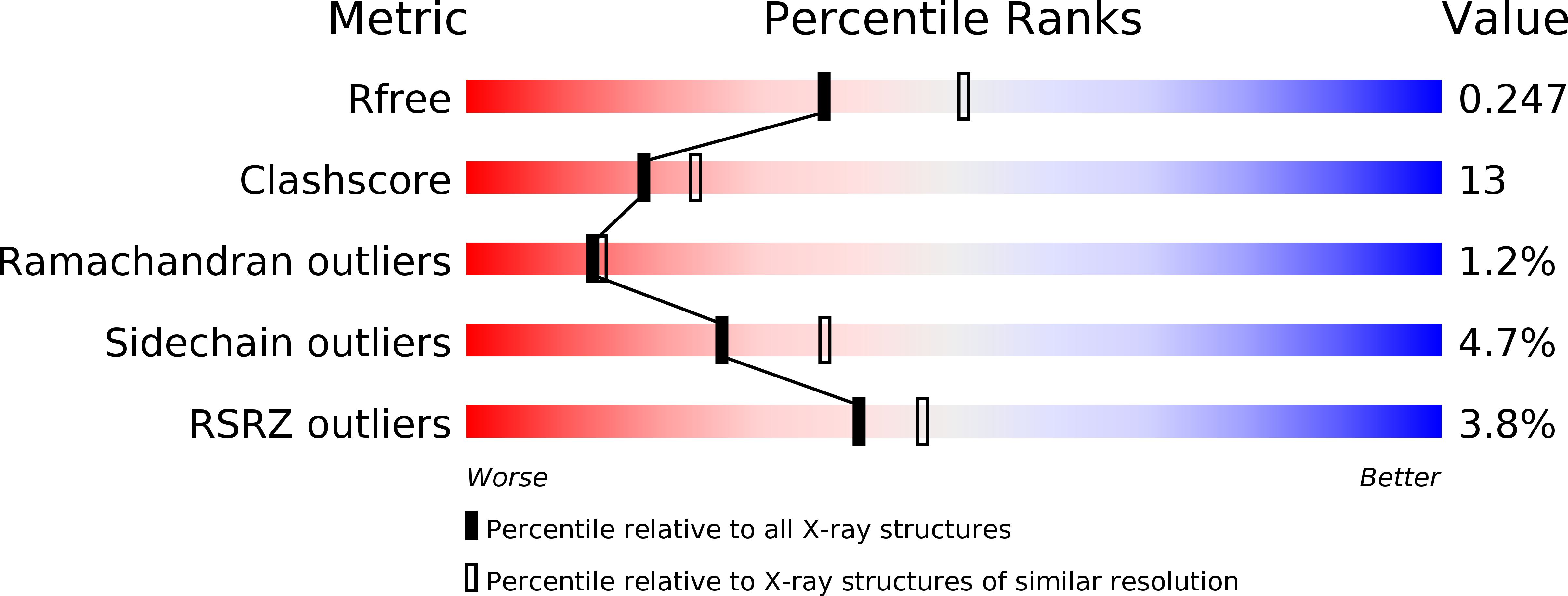
Resolution:
2.30 Å
R-Value Free:
0.24
R-Value Work:
0.18
R-Value Observed:
0.18
Space Group:
P 21 21 21
Macromolecular Entities
Polymer Type:polypeptide(L)
Molecule:EhpF
Gene (Uniprot):ehpF
Chain IDs:A, B
Chain Length:369
Number of Molecules:2
Biological Source:Pantoea agglomerans
Modified Residue
Compound ID Chain ID Parent Comp ID Details 2D Image
MSE A MET SELENOMETHIONINE
Primary Citation
Structure of the D-alanylgriseoluteic acid biosynthetic protein EhpF, an atypical member of the ANL superfamily of adenylating enzymes.
Acta Crystallogr. D Biol. Crystallogr. 66 664 672 (2010)
PMID: 20516619 DOI: 10.1107/S0907444910008425

Abstact

The structure of EhpF, a 41 kDa protein that functions in the biosynthetic pathway leading to the broad-spectrum antimicrobial compound D-alanylgriseoluteic acid (AGA), is reported. A cluster of approximately 16 genes, including ehpF, located on a 200 kbp plasmid native to certain strains of Pantoea agglomerans encodes the proteins that are required for the conversion of chorismic acid to AGA. Phenazine-1,6-dicarboxylate has been identified as an intermediate in AGA biosynthesis and deletion of ehpF results in accumulation of this compound in vivo. The crystallographic data presented here reveal that EhpF is an atypical member of the acyl-CoA synthase or ANL superfamily of adenylating enzymes. These enzymes typically catalyze two-step reactions involving adenylation of a carboxylate substrate followed by transfer of the substrate from AMP to coenzyme A or another phosphopantetheine. EhpF is distinguished by the absence of the C-terminal domain that is characteristic of enzymes from this family and is involved in phosphopantetheine binding and in the second half of the canonical two-step reaction that is typically observed. Based on the structure of EhpF and a bioinformatic analysis, it is proposed that EhpF and EhpG convert phenazine-1,6-dicarboxylate to 6-formylphenazine-1-carboxylate via an adenylyl intermediate.

Legend

Protein

Chemical

Disease

Primary Citation of related structures