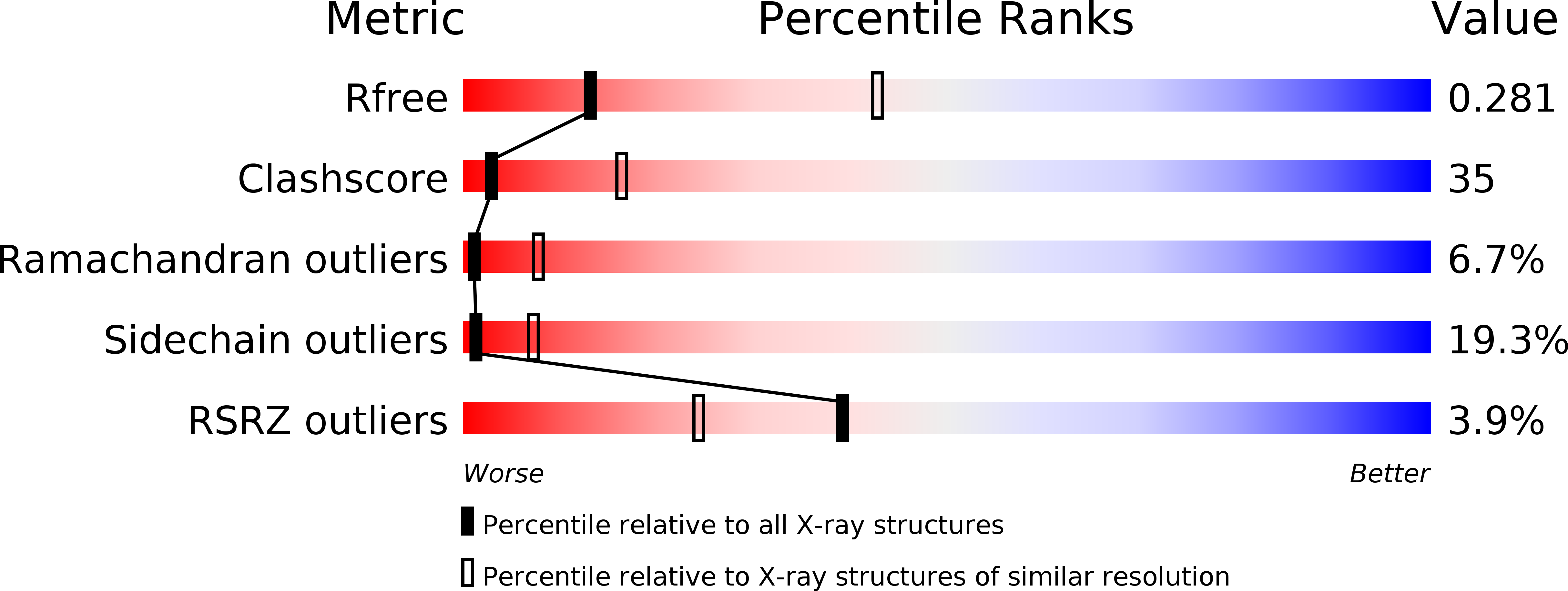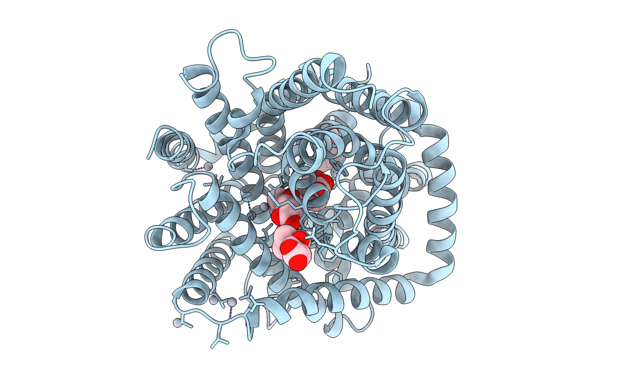
3HFX image
Deposition Date 2009-05-13
Release Date 2010-03-31
Last Version Date 2024-03-20
Entry Detail
PDB ID:
3HFX
Title:
Crystal structure of carnitine transporter
Biological Source:
Source Organism:
Host Organism:
Method Details:
Experimental Method:
Resolution:
3.15 Å
R-Value Free:
0.28
R-Value Work:
0.26
Space Group:
P 63
Macromolecular Entities
Structures with similar UniProt ID
Protein Blast
Polymer Type:polypeptide(L)
Molecule:L-carnitine/gamma-butyrobetaine antiporter
Gene (Uniprot):caiT
Chain IDs:A
Chain Length:504
Number of Molecules:1
Biological Source:Escherichia coli
Primary Citation
Crystal structure of the carnitine transporter and insights into the antiport mechanism
Nat.Struct.Mol.Biol. 17 492 496 (2010)
PMID: 20357772 DOI: 10.1038/nsmb.1788

Abstact

CaiT is a membrane antiporter that catalyzes the exchange of L-carnitine with gamma-butyrobetaine across the Escherichia coli membrane. To obtain structural insights into the antiport mechanism, we solved the crystal structure of CaiT at a resolution of 3.15 A. We crystallized CaiT as a homotrimer complex, in which each protomer contained 12 transmembrane helices and 4 l-carnitine molecules outlining the transport pathway across the membrane. Mutagenesis studies revealed a primary binding site at the center of the protein and a secondary substrate-binding site at the bottom of the intracellular vestibule. These results, together with the insights obtained from structural comparison with structurally homologous transporters, provide mechanistic insights into the association between substrate translocation and the conformational changes of CaiT.

Legend

Protein

Chemical

Disease

Primary Citation of related structures