3HFS image
Deposition Date 2009-05-12
Release Date 2009-09-01
Last Version Date 2023-09-06
Entry Detail
PDB ID:
3HFS
Keywords:
Title:
Structure of apo anthocyanidin reductase from vitis vinifera
Biological Source:
Source Organism:
Vitis vinifera (Taxon ID: 29760)
Host Organism:
Method Details:
Experimental Method:
Resolution:
3.17 Å
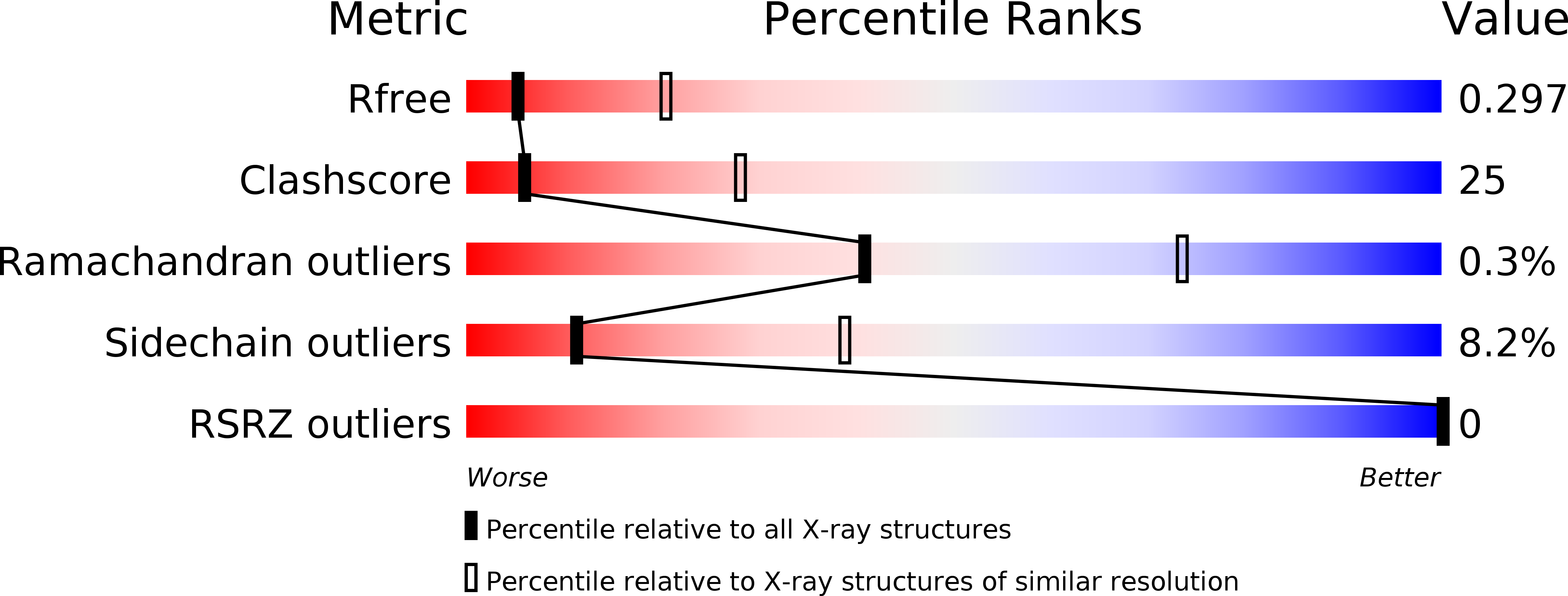
R-Value Free:
0.29
R-Value Work:
0.22
R-Value Observed:
0.22
Space Group:
P 1 21 1
Macromolecular Entities
Structures with similar UniProt ID
Protein Blast
Polymer Type:polypeptide(L)
Molecule:Anthocyanidin reductase
Gene (Uniprot):ANR
Chain IDs:A, B
Chain Length:338
Number of Molecules:2
Biological Source:Vitis vinifera
Ligand Molecules
Primary Citation
Structure and epimerase activity of anthocyanidin reductase from Vitis vinifera.
Acta Crystallogr.,Sect.D 65 989 1000 (2009)
PMID: 19690377 DOI: 10.1107/S0907444909025013

Abstact

Together with leucoanthocyanidin reductase, anthocyanidin reductase (ANR) is one of the two enzymes of the flavonoid-biosynthesis pathway that produces the flavan-3-ol monomers required for the formation of proanthocyanidins or condensed tannins. It has been shown to catalyse the double reduction of anthocyanidins to form 2R,3R-flavan-3-ols, which can be further transformed to the 2S,3R isomers by non-enzymatic epimerization. ANR from grape (Vitis vinifera) was expressed in Escherichia coli and purified. Unexpectedly, RP-HPLC, LC-MS and NMR experiments clearly established that the enzyme produces a 50:50 mixture of 2,3-cis and 2,3-trans flavan-3-ols which have been identified by chiral chromatography to be 2S,3S- and 2S,3R-flavan-3-ols, i.e. the naturally rare (+)-epicatechin and (-)-catechin, when cyanidin is used as the substrate of the reaction. The first three-dimensional structure of ANR is described at a resolution of 2.2 A and explains the inactivity of the enzyme in the presence of high salt concentrations.

Legend

Protein

Chemical

Disease

Primary Citation of related structures