3HDL image
Deposition Date 2009-05-07
Release Date 2009-11-24
Last Version Date 2024-10-30
Entry Detail
PDB ID:
3HDL
Keywords:
Title:
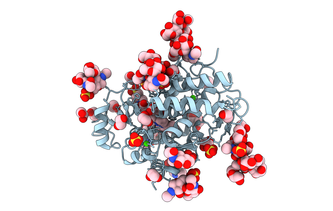
Crystal Structure of Highly Glycosylated Peroxidase from Royal Palm Tree
Biological Source:
Source Organism:
Method Details:
Experimental Method:
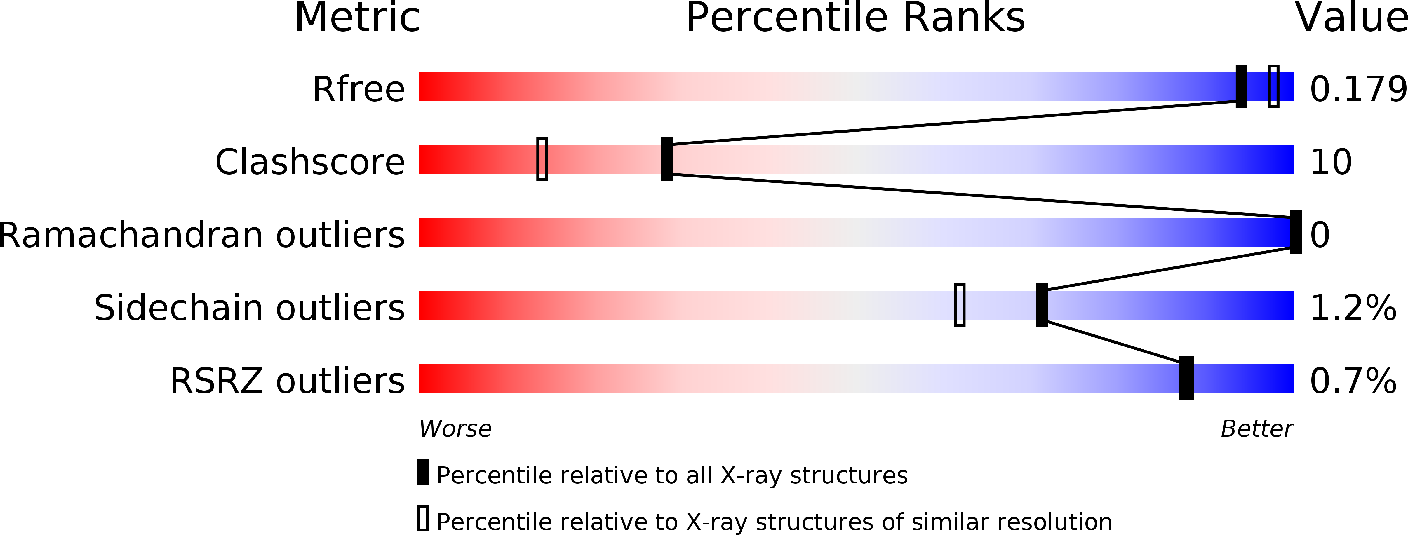
Resolution:
1.85 Å
R-Value Free:
0.18
R-Value Work:
0.17
R-Value Observed:
0.17
Space Group:
P 31 2 1
Macromolecular Entities
Polymer Type:polypeptide(L)
Molecule:Royal Palm Tree Peroxidase
Chain IDs:A
Chain Length:304
Number of Molecules:1
Biological Source:Roystonea regia
Modified Residue
Compound ID Chain ID Parent Comp ID Details 2D Image
ASN A ASN GLYCOSYLATION SITE
Primary Citation
Crystal structure and statistical coupling analysis of highly glycosylated peroxidase from royal palm tree (Roystonea regia).
J.Struct.Biol. 169 226 242 (2010)
PMID: 19854274 DOI: 10.1016/j.jsb.2009.10.009

Abstact

Royal palm tree peroxidase (RPTP) is a very stable enzyme in regards to acidity, temperature, H(2)O(2), and organic solvents. Thus, RPTP is a promising candidate for developing H(2)O(2)-sensitive biosensors for diverse applications in industry and analytical chemistry. RPTP belongs to the family of class III secretory plant peroxidases, which include horseradish peroxidase isozyme C, soybean and peanut peroxidases. Here we report the X-ray structure of native RPTP isolated from royal palm tree (Roystonea regia) refined to a resolution of 1.85A. RPTP has the same overall folding pattern of the plant peroxidase superfamily, and it contains one heme group and two calcium-binding sites in similar locations. The three-dimensional structure of RPTP was solved for a hydroperoxide complex state, and it revealed a bound 2-(N-morpholino) ethanesulfonic acid molecule (MES) positioned at a putative substrate-binding secondary site. Nine N-glycosylation sites are clearly defined in the RPTP electron-density maps, revealing for the first time conformations of the glycan chains of this highly glycosylated enzyme. Furthermore, statistical coupling analysis (SCA) of the plant peroxidase superfamily was performed. This sequence-based method identified a set of evolutionarily conserved sites that mapped to regions surrounding the heme prosthetic group. The SCA matrix also predicted a set of energetically coupled residues that are involved in the maintenance of the structural folding of plant peroxidases. The combination of crystallographic data and SCA analysis provides information about the key structural elements that could contribute to explaining the unique stability of RPTP.

Legend

Protein

Chemical

Disease

Primary Citation of related structures