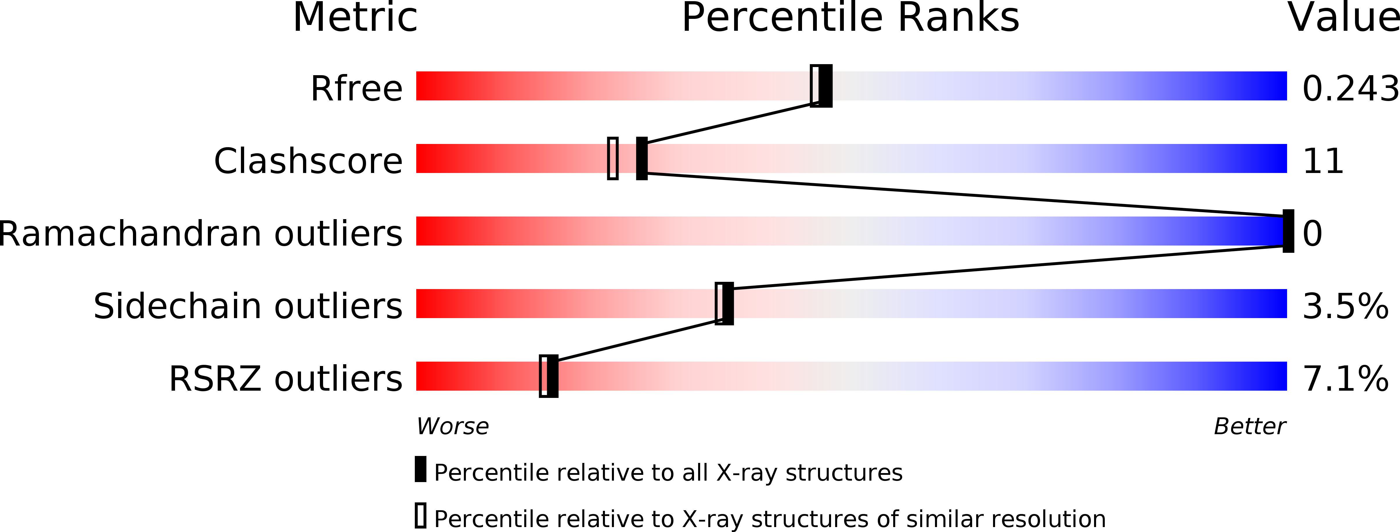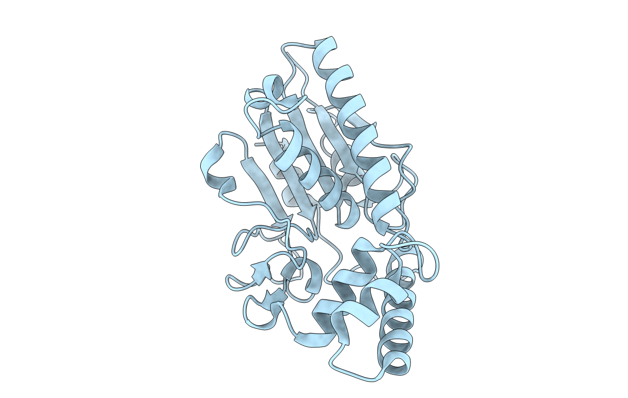
3HC7 image
Deposition Date 2009-05-05
Release Date 2009-07-21
Last Version Date 2024-02-21
Entry Detail
PDB ID:
3HC7
Keywords:
Title:
Crystal structure of lysin B from Mycobacteriophage D29
Biological Source:
Source Organism:
Host Organism:
Method Details:
Experimental Method:
Resolution:
2.00 Å
R-Value Free:
0.25
R-Value Work:
0.20
Space Group:
P 43 21 2
Macromolecular Entities
Polymer Type:polypeptide(L)
Molecule:Gene 12 protein
Gene (Uniprot):12
Chain IDs:A
Chain Length:254
Number of Molecules:1
Biological Source:Mycobacterium phage D29
Primary Citation
Mycobacteriophage Lysin B is a novel mycolylarabinogalactan esterase
Mol.Microbiol. 73 367 381 (2009)
PMID: 19555454 DOI: 10.1111/j.1365-2958.2009.06775.x

Abstact

Mycobacteriophages encounter a unique problem among phages of Gram-positive bacteria, in that lysis must not only degrade the peptidoglycan layer but also circumvent a mycolic acid-rich outer membrane covalently attached to the arabinogalactan-peptidoglycan complex. Mycobacteriophages accomplish this by producing two lysis enzymes, Lysin A (LysA) that hydrolyses peptidoglycan, and Lysin B (LysB), a novel mycolylarabinogalactan esterase, that cleaves the mycolylarabinogalactan bond to release free mycolic acids. The D29 LysB structure shows an alpha/beta hydrolase organization with a catalytic triad common to cutinases, but which contains an additional four-helix domain implicated in the binding of lipid substrates. Whereas LysA is essential for mycobacterial lysis, a Giles DeltalysB mutant mycobacteriophage is viable, but defective in the normal timing, progression and completion of host cell lysis. We propose that LysB facilitates lysis by compromising the integrity of the mycobacterial outer membrane linkage to the arabinogalactan-peptidoglycan layer.

Legend

Protein

Chemical

Disease

Primary Citation of related structures