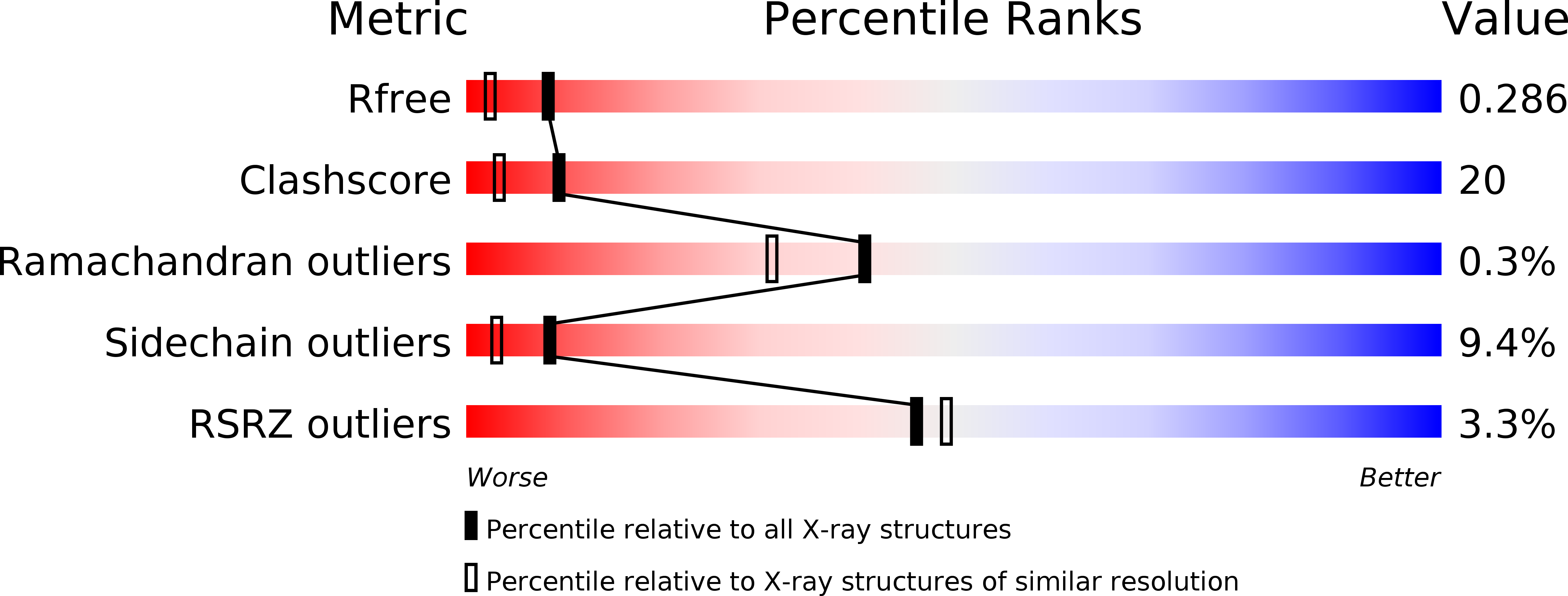
3HBR image
Deposition Date 2009-05-05
Release Date 2009-06-23
Last Version Date 2023-11-22
Entry Detail
PDB ID:
3HBR
Keywords:
Title:
Crystal structure of OXA-48 beta-lactamase
Biological Source:
Source Organism:
Host Organism:
Method Details:
Experimental Method:
Resolution:
1.90 Å
R-Value Free:
0.28
R-Value Work:
0.21
R-Value Observed:
0.22
Space Group:
P 1 21 1
Macromolecular Entities
Structures with similar UniProt ID
Protein Blast
Polymer Type:polypeptide(L)
Molecule:OXA-48
Gene (Uniprot):OXA-48
Chain IDs:A, B, C, D
Chain Length:265
Number of Molecules:4
Biological Source:Klebsiella pneumoniae
Modified Residue
Compound ID Chain ID Parent Comp ID Details 2D Image
KCX A LYS LYSINE NZ-CARBOXYLIC ACID
Ligand Molecules
Primary Citation
Crystal structure of the OXA-48 beta-lactamase reveals mechanistic diversity among class D carbapenemases.
Chem.Biol. 16 540 547 (2009)
PMID: 19477418 DOI: 10.1016/j.chembiol.2009.04.010

Abstact

Carbapenem-hydrolyzing class D beta-lactamases (CHDLs) are enzymes found in important Gram-negative pathogens (mainly Acinetobacter baumannii and Enterobacteriaceae) that confer resistance to beta-lactam antibiotics, and notably carbapenems. The crystal structure of the OXA-48 carbapenemase was determined at pH 7.5 and at a resolution of 1.9 A. Surprisingly, and by contrast with OXA-24, the only other CHDL of known crystal structure, the structure of OXA-48 was similar to OXA-10, an enzyme devoid of carbapenemase activity, indicating that the hydrolysis of these compounds could depend on subtle changes in the active site region. Moreover, the active site groove of OXA-48 was different from that of OXA-24 in shape, dimensions, and charge distribution. Molecular dynamics pointed to the functional relevance of residues located in or close to the beta5-beta6 loop and allowed us to propose a mechanism for carbapenem hydrolysis by OXA-48.

Legend

Protein

Chemical

Disease

Primary Citation of related structures