Deposition Date
2009-05-04
Release Date
2009-06-23
Last Version Date
2024-11-27
Entry Detail
PDB ID:
3HB3
Keywords:
Title:
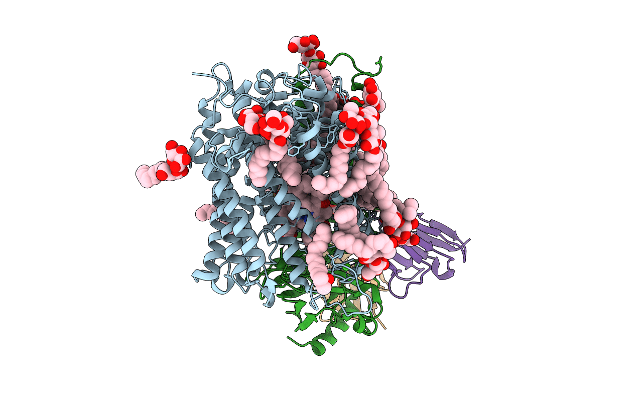
High resolution crystal structure of Paracoccus denitrificans cytochrome c oxidase
Biological Source:
Source Organism(s):
Paracoccus denitrificans (Taxon ID: 266)
Mus musculus (Taxon ID: 10090)
Mus musculus (Taxon ID: 10090)
Expression System(s):
Method Details:
Experimental Method:
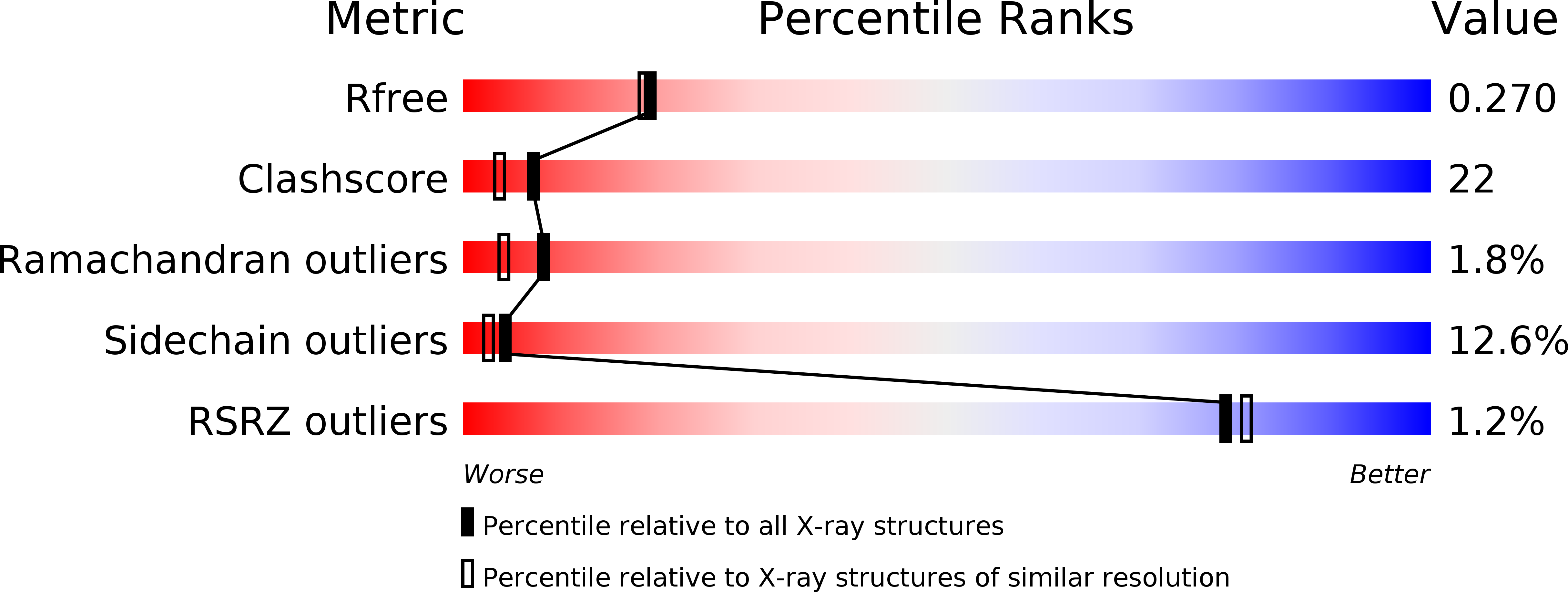
Resolution:
2.25 Å
R-Value Free:
0.27
R-Value Work:
0.21
R-Value Observed:
0.22
Space Group:
P 21 21 21