3HAP image
Deposition Date 2009-05-02
Release Date 2009-09-22
Last Version Date 2024-10-30
Entry Detail
PDB ID:
3HAP
Title:
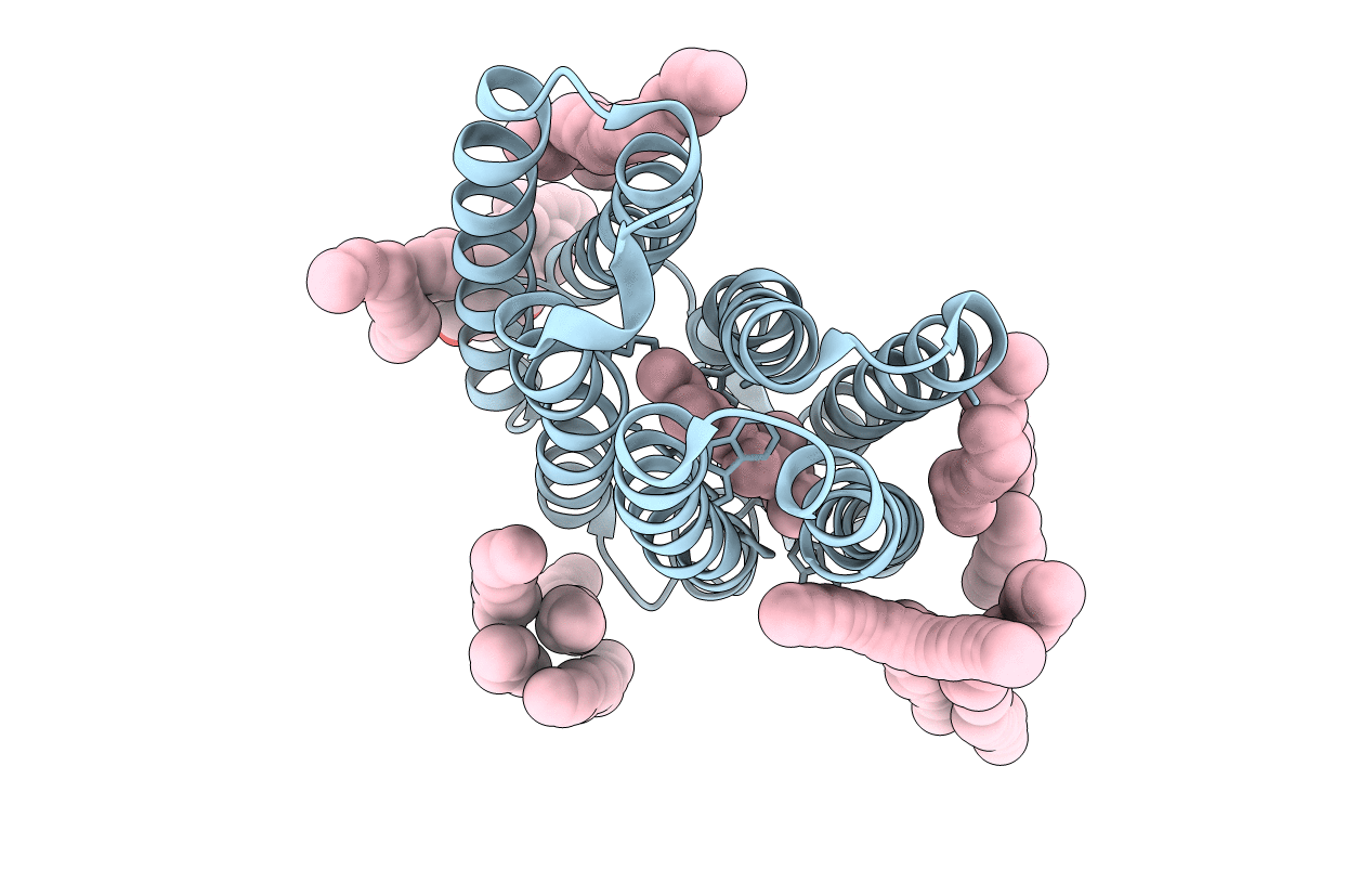
Crystal structure of bacteriorhodopsin mutant L111A crystallized from bicelles
Biological Source:
Source Organism:
Method Details:
Experimental Method:
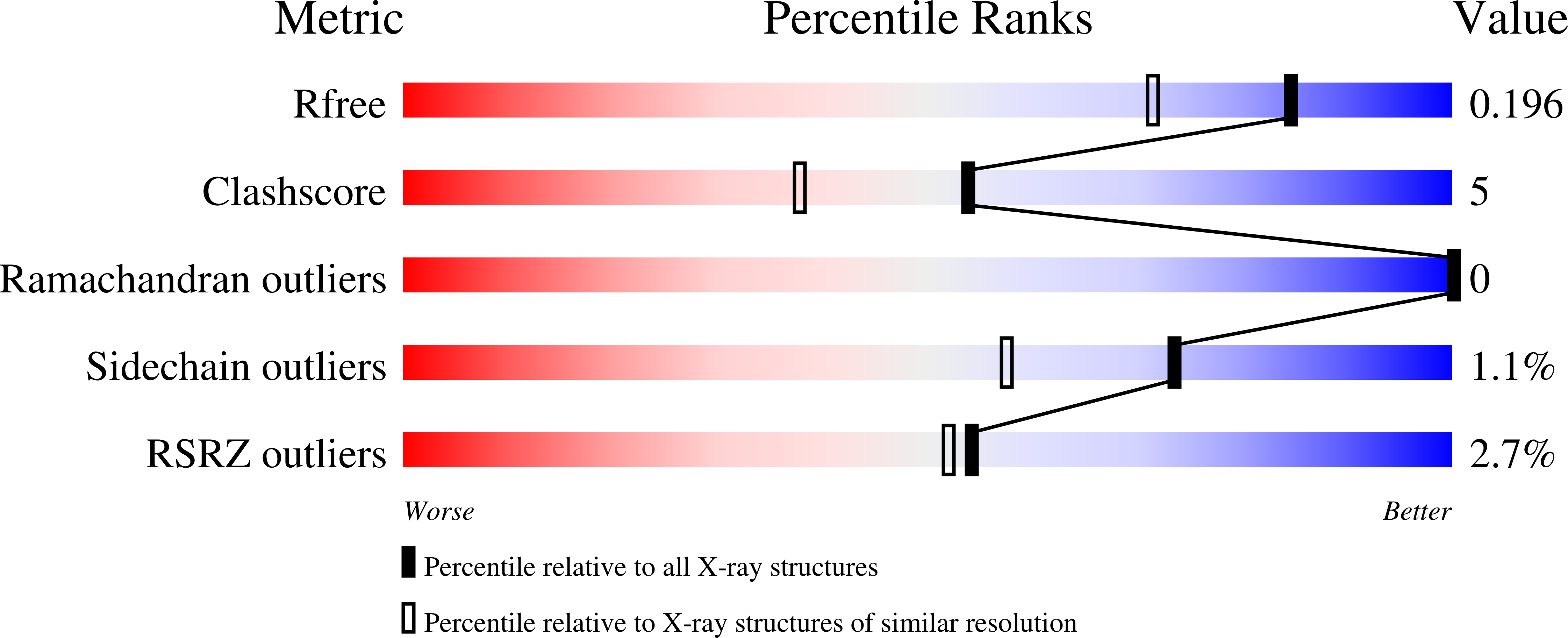
Resolution:
1.60 Å
R-Value Free:
0.19
R-Value Work:
0.16
R-Value Observed:
0.16
Space Group:
C 2 2 21
Macromolecular Entities
Structures with similar UniProt ID
Protein Blast
Polymer Type:polypeptide(L)
Molecule:Bacteriorhodopsin
Gene (Uniprot):bop
Mutagens:L111A
Chain IDs:A
Chain Length:249
Number of Molecules:1
Biological Source:Halobacterium salinarum
Primary Citation
Similar energetic contributions of packing in the core of membrane and water-soluble proteins.
J.Am.Chem.Soc. 131 10846 10847 (2009)
PMID: 19603754 DOI: 10.1021/ja904711k

Abstact

A major driving force for water-soluble protein folding is the hydrophobic effect, but membrane proteins cannot make use of this stabilizing contribution in the apolar core of the bilayer. It has been proposed that membrane proteins compensate by packing more efficiently. We therefore investigated packing contributions experimentally by observing the energetic and structural consequences of cavity creating mutations in the core of a membrane protein. We observed little difference in the packing energetics of water and membrane soluble proteins. Our results imply that other mechanisms are employed to stabilize the structure of membrane proteins.

Legend

Protein

Chemical

Disease

Primary Citation of related structures