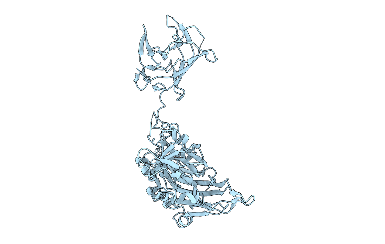
3HAG image
Deposition Date 2009-05-01
Release Date 2009-09-01
Last Version Date 2024-04-03
Entry Detail
PDB ID:
3HAG
Keywords:
Title:
Crystal structure of the Hepatitis E Virus-like Particle
Biological Source:
Source Organism:
Host Organism:
Method Details:
Experimental Method:
Resolution:
3.50 Å
R-Value Free:
0.28
R-Value Work:
0.27
Space Group:
P 63
Macromolecular Entities
Polymer Type:polypeptide(L)
Molecule:Capsid protein
Chain IDs:A
Chain Length:504
Number of Molecules:1
Biological Source:Hepatitis E virus type 4
Ligand Molecules
Primary Citation
Structure of the hepatitis E virus-like particle suggests mechanisms for virus assembly and receptor binding.
Proc.Natl.Acad.Sci.USA 106 12992 12997 (2009)
PMID: 19622744 DOI: 10.1073/pnas.0904848106

Abstact

Hepatitis E virus (HEV), a small, non-enveloped RNA virus in the family Hepeviridae, is associated with endemic and epidemic acute viral hepatitis in developing countries. Our 3.5-A structure of a HEV-like particle (VLP) shows that each capsid protein contains 3 linear domains that form distinct structural elements: S, the continuous capsid; P1, 3-fold protrusions; and P2, 2-fold spikes. The S domain adopts a jelly-roll fold commonly observed in small RNA viruses. The P1 and P2 domains both adopt beta-barrel folds. Each domain possesses a potential polysaccharide-binding site that may function in cell-receptor binding. Sugar binding to P1 at the capsid protein interface may lead to capsid disassembly and cell entry. Structural modeling indicates that native T = 3 capsid contains flat dimers, with less curvature than those of T = 1 VLP. Our findings significantly advance the understanding of HEV molecular biology and have application to the development of vaccines and antiviral medications.

Legend

Protein

Chemical

Disease

Primary Citation of related structures