3H9Z image
Deposition Date 2009-04-30
Release Date 2009-09-08
Last Version Date 2024-10-16
Entry Detail
PDB ID:
3H9Z
Keywords:
Title:
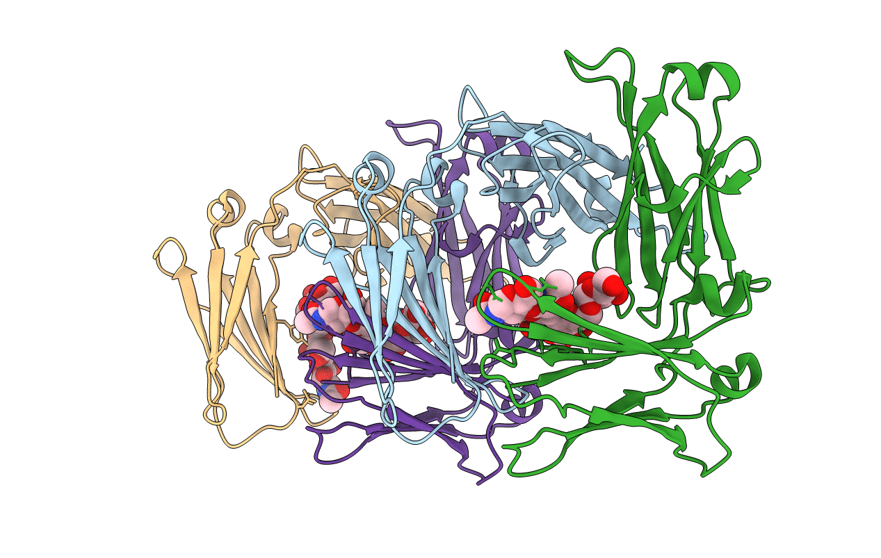
Crystal structure of the IgE-Fc3-4 domains
Biological Source:
Source Organism:
Homo sapiens (Taxon ID: 9606)
Host Organism:
Method Details:
Experimental Method:
Resolution:
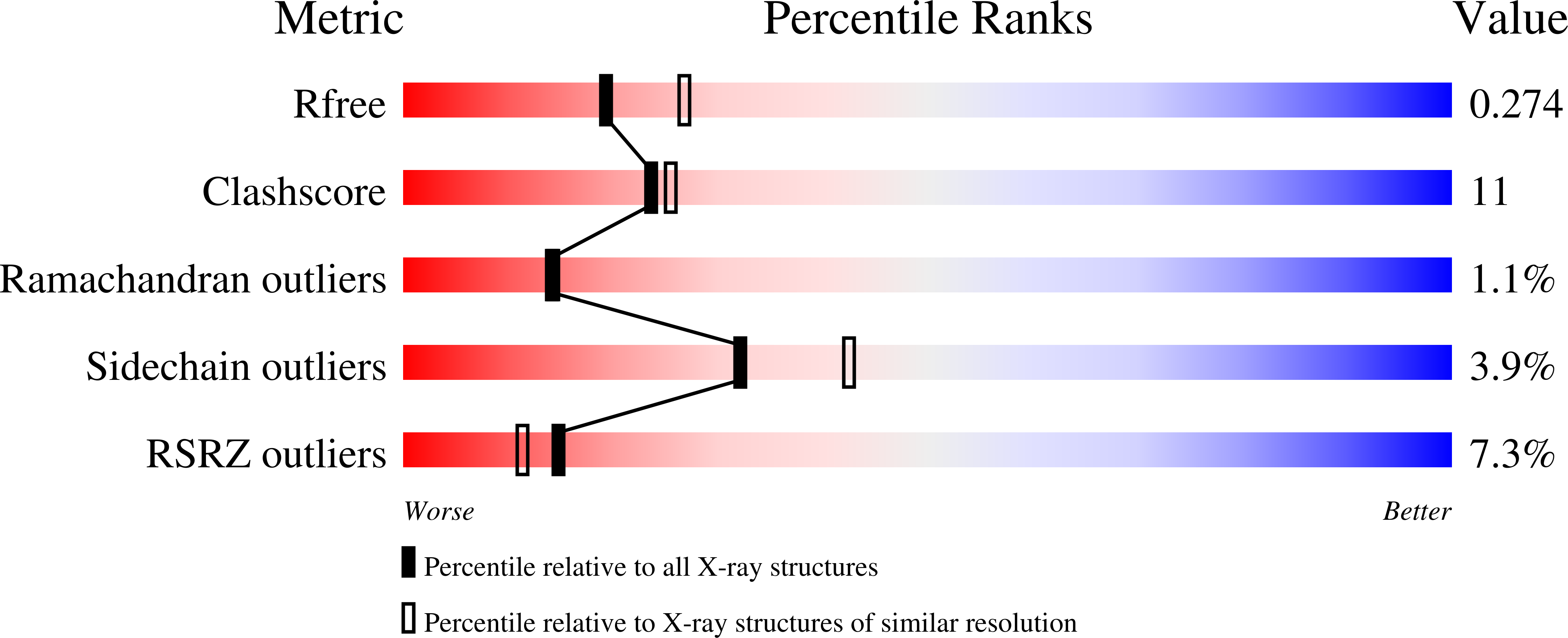
2.45 Å
R-Value Free:
0.27
R-Value Work:
0.22
R-Value Observed:
0.23
Space Group:
P 1 21 1
Macromolecular Entities
Structures with similar UniProt ID
Protein Blast
Polymer Type:polypeptide(L)
Molecule:Ig epsilon chain C region
Gene (Uniprot):IGHE
Mutagens:N252Q, N264Q
Chain IDs:A, B, C, D
Chain Length:223
Number of Molecules:4
Biological Source:Homo sapiens
Modified Residue
Compound ID Chain ID Parent Comp ID Details 2D Image
ASN B ASN GLYCOSYLATION SITE
Ligand Molecules
Primary Citation
Conformational flexibility in immunoglobulin E-Fc 3-4 revealed in multiple crystal forms.
J.Mol.Biol. 393 176 190 (2009)
PMID: 19682998 DOI: 10.1016/j.jmb.2009.08.012

Abstact

The structure of immunoglobulin E (IgE)-Fc(3-4) has been solved in three new crystal forms, providing 13 snapshots of the Fc conformation and revealing a diverse range of open-closed motions among subunit chains and dimers. A more detailed analysis of the open-to-closed motion of IgE-Fc(3-4) was possible with so many structures, and the new structures allow a more thorough examination of the flexibility of IgE-Fc and its implications for receptor binding. The existence of a hydrophobic pocket at the elbow region of the Fc appears to be conformation dependent and suggests a means of regulating the IgE-Fc conformation (and potentially receptor binding) with small molecules.

Legend

Protein

Chemical

Disease

Primary Citation of related structures