3H9R image
Deposition Date 2009-04-30
Release Date 2009-06-02
Last Version Date 2023-09-06
Entry Detail
PDB ID:
3H9R
Title:
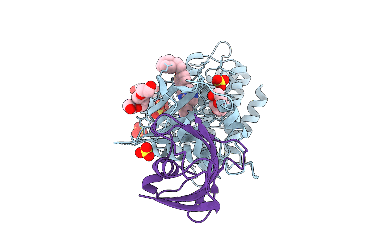
Crystal structure of the kinase domain of type I activin receptor (ACVR1) in complex with FKBP12 and dorsomorphin
Biological Source:
Source Organism:
Homo sapiens (Taxon ID: 9606)
Host Organism:
Method Details:
Experimental Method:
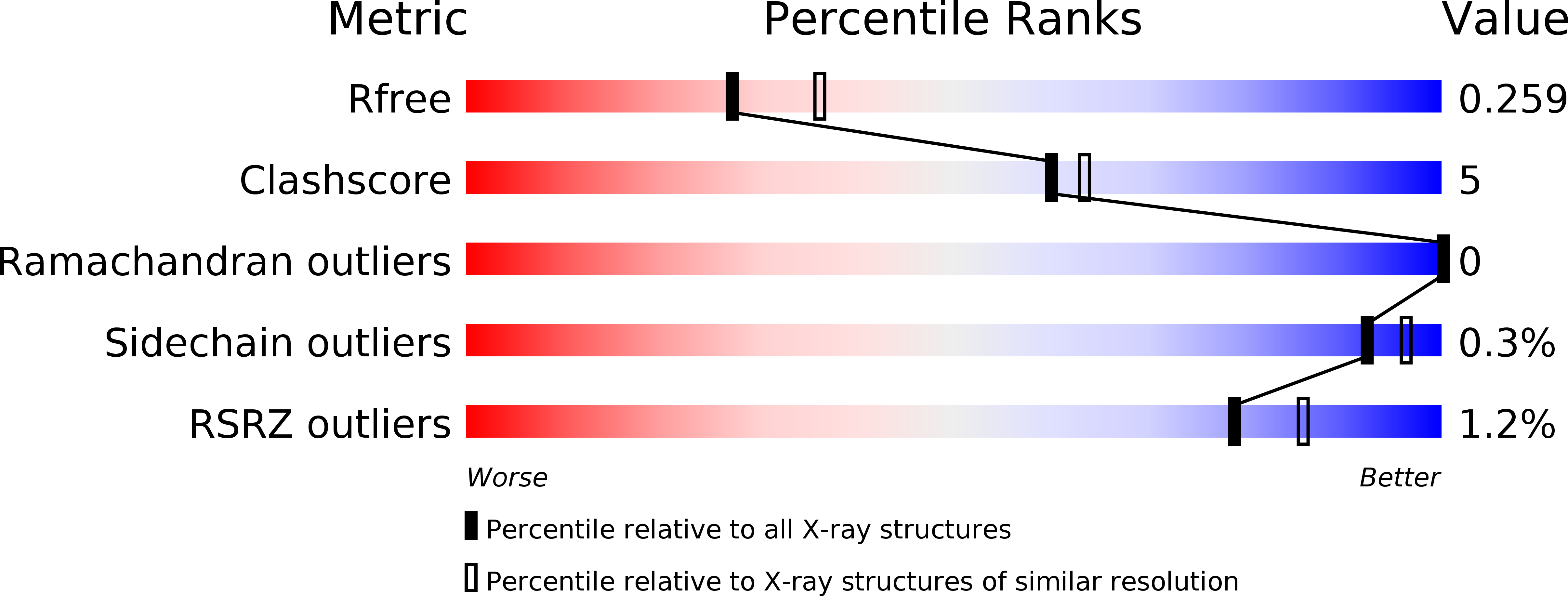
Resolution:
2.35 Å
R-Value Free:
0.25
R-Value Work:
0.18
R-Value Observed:
0.19
Space Group:
P 21 21 21
Macromolecular Entities
Structures with similar UniProt ID
Protein Blast
Polymer Type:polypeptide(L)
Molecule:Activin receptor type-1
Gene (Uniprot):ACVR1
Chain IDs:A
Chain Length:330
Number of Molecules:1
Biological Source:Homo sapiens
Structures with similar UniProt ID
Protein Blast
Polymer Type:polypeptide(L)
Molecule:Peptidyl-prolyl cis-trans isomerase FKBP1A
Gene (Uniprot):FKBP1A
Chain IDs:B
Chain Length:109
Number of Molecules:1
Biological Source:Homo sapiens
Primary Citation
Structure of the Bone Morphogenetic Protein Receptor ALK2 and Implications for Fibrodysplasia Ossificans Progressiva.
J.Biol.Chem. 287 36990 36998 (2012)
PMID: 22977237 DOI: 10.1074/jbc.M112.365932

Abstact

Bone morphogenetic protein (BMP) receptor kinases are tightly regulated to control development and tissue homeostasis. Mutant receptor kinase domains escape regulation leading to severely degenerative diseases and represent an important therapeutic target. Fibrodysplasia ossificans progressiva (FOP) is a rare but devastating disorder of extraskeletal bone formation. FOP-associated mutations in the BMP receptor ALK2 reduce binding of the inhibitor FKBP12 and promote leaky signaling in the absence of ligand. To establish structural mechanisms of receptor regulation and to address the effects of FOP mutation, we determined the crystal structure of the cytoplasmic domain of ALK2 in complex with the inhibitors FKBP12 and dorsomorphin. FOP mutations break critical interactions that stabilize the inactive state of the kinase, thereby facilitating structural rearrangements that diminish FKBP12 binding and promote the correct positioning of the glycine-serine-rich loop and αC helix for kinase activation. The balance of these effects accounts for the comparable activity of R206H and L196P. Kinase activation in the clinically benign mutant L196P is far weaker than R206H but yields equivalent signals due to the stronger interaction of FKBP12 with R206H. The presented ALK2 structure offers a valuable template for the further design of specific inhibitors of BMP signaling.

Legend

Protein

Chemical

Disease

Primary Citation of related structures
Feedback Form
Name
Email
Institute
Feedback