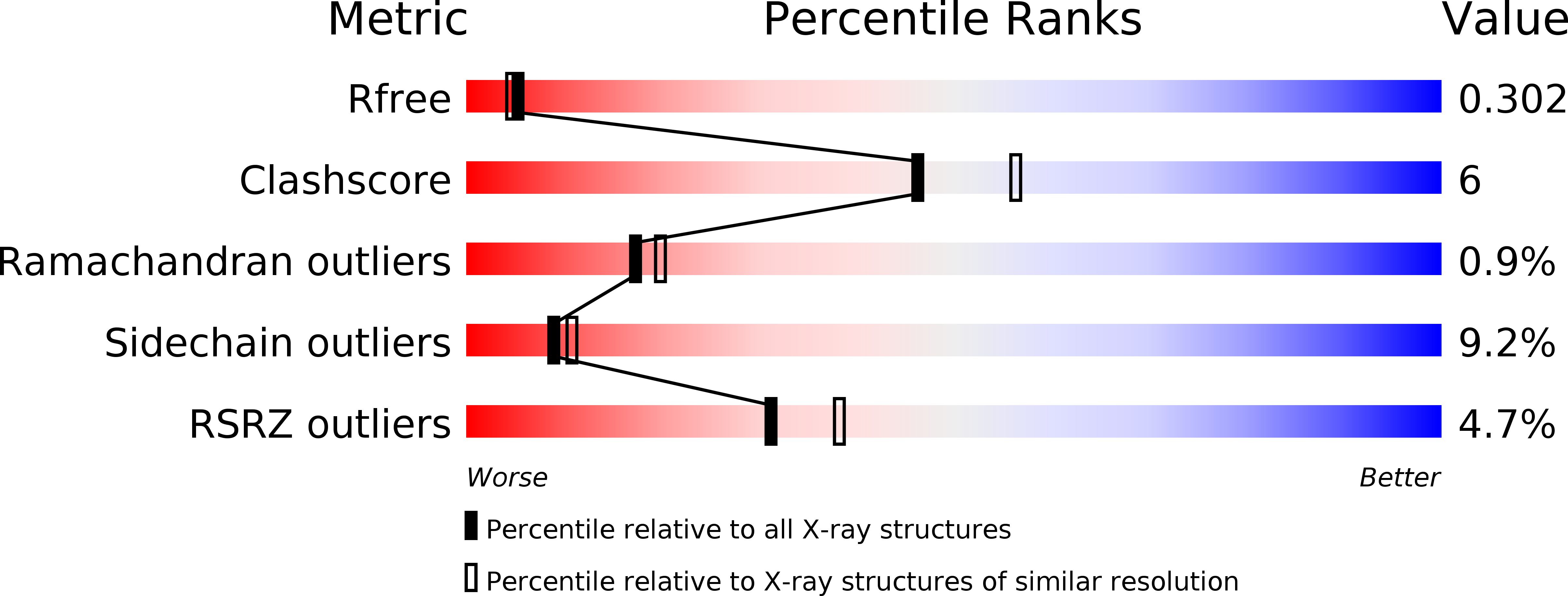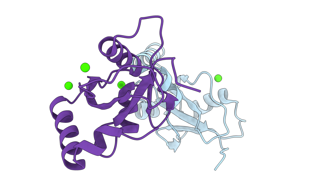
3H9D image
Deposition Date 2009-04-30
Release Date 2009-10-06
Last Version Date 2024-02-21
Entry Detail
PDB ID:
3H9D
Title:
Crystal Structure of Trypanosoma brucei ATG8
Biological Source:
Source Organism:
Host Organism:
Method Details:
Experimental Method:
Resolution:
2.30 Å
R-Value Free:
0.30
R-Value Work:
0.23
R-Value Observed:
0.23
Space Group:
P 21 21 21
Macromolecular Entities
Structures with similar UniProt ID
Protein Blast
Polymer Type:polypeptide(L)
Molecule:Microtubule-associated protein 1A/1B, light chain 3, putative
Gene (Uniprot):Tb07.10C21.40
Chain IDs:A, B
Chain Length:119
Number of Molecules:2
Biological Source:Trypanosoma brucei
Ligand Molecules
Primary Citation
Trypanosoma brucei ATG8: structural insights into autophagic-like mechanisms in protozoa.
Autophagy 5 1085 1091 (2009)
PMID: 19736525

Abstact

Bioinformatic searches of genome databases revealed that the number of autophagy-related genes (ATG) is considerably lower in trypanosomes than in higher eukaryotes and even in yeast. This raises the question of whether autophagy in this protozoan parasite is more primitive and represents a rudimentary paradigm due to its early branching off the evolutionary tree. We here present the crystal structure of TbATG8B. This molecule (MW 13.7 kDa) belongs to the ubiquitin-like proteins showing the typical ubiquitin fold and strong sequence homology to LC3, the human homologue. Due to its characteristic folding, it should readily bind to TbATG4.1 for being processed. This presumption was tested by molecular modeling approaches, docking TbATG8B to a homology model of TbATG4.1. Although exchanges of several amino acids are evident from sequence comparisons, the overall structure seems very much alike and the necessary catalytic triad (C-D-H) is well conserved in TbATG4.1. Thus membrane formation during appearance of the autophagic bodies seems very similar in trypanosomes and their higher eukaryotic counterpart.

Legend

Protein

Chemical

Disease

Primary Citation of related structures