Deposition Date
2009-04-29
Release Date
2009-09-29
Last Version Date
2024-03-20
Entry Detail
PDB ID:
3H8D
Keywords:
Title:
Crystal structure of Myosin VI in complex with Dab2 peptide
Biological Source:
Source Organism(s):
Mus musculus (Taxon ID: 10090)
Rattus norvegicus (Taxon ID: 10116)
Rattus norvegicus (Taxon ID: 10116)
Expression System(s):
Method Details:
Experimental Method:
Resolution:
2.20 Å
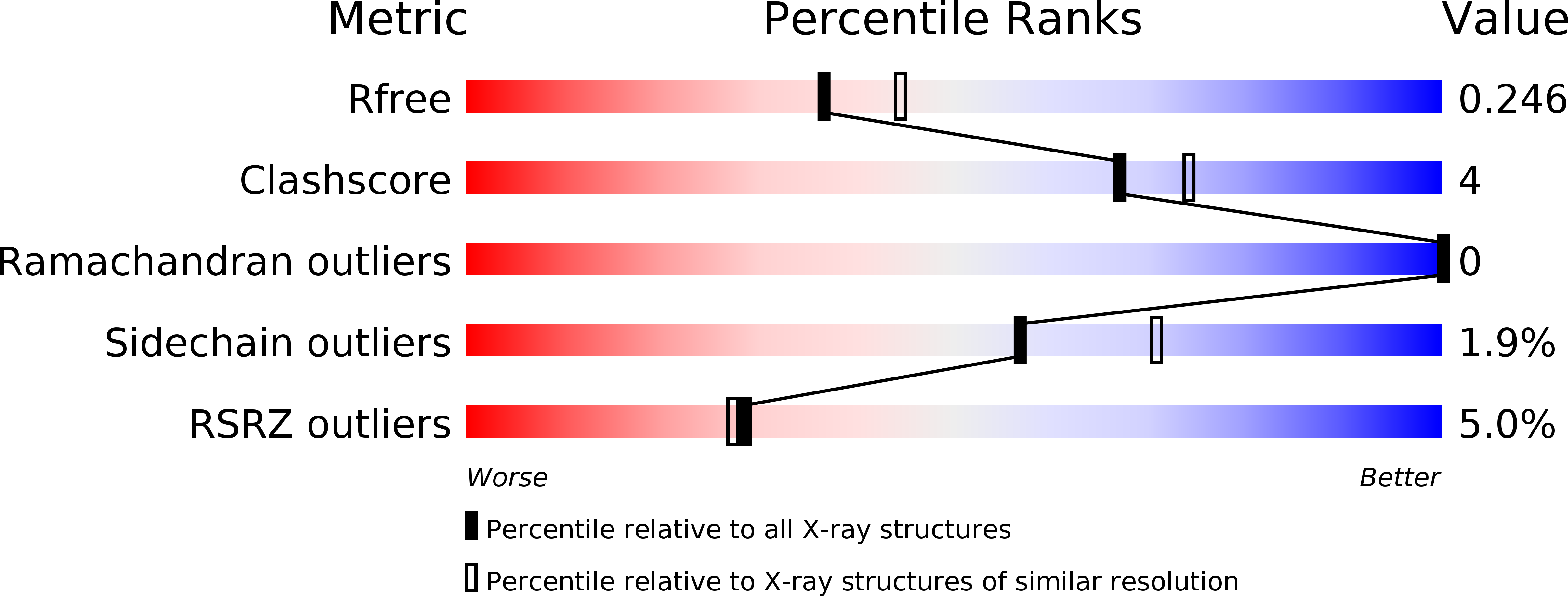
R-Value Free:
0.25
R-Value Work:
0.18
R-Value Observed:
0.19
Space Group:
P 1 21 1