3H8C image
Deposition Date 2009-04-29
Release Date 2009-10-20
Last Version Date 2024-10-16
Entry Detail
PDB ID:
3H8C
Keywords:
Title:
A combined crystallographic and molecular dynamics study of cathepsin-L retro-binding inhibitors (compound 14)
Biological Source:
Source Organism:
Homo sapiens (Taxon ID: 9606)
Method Details:
Experimental Method:
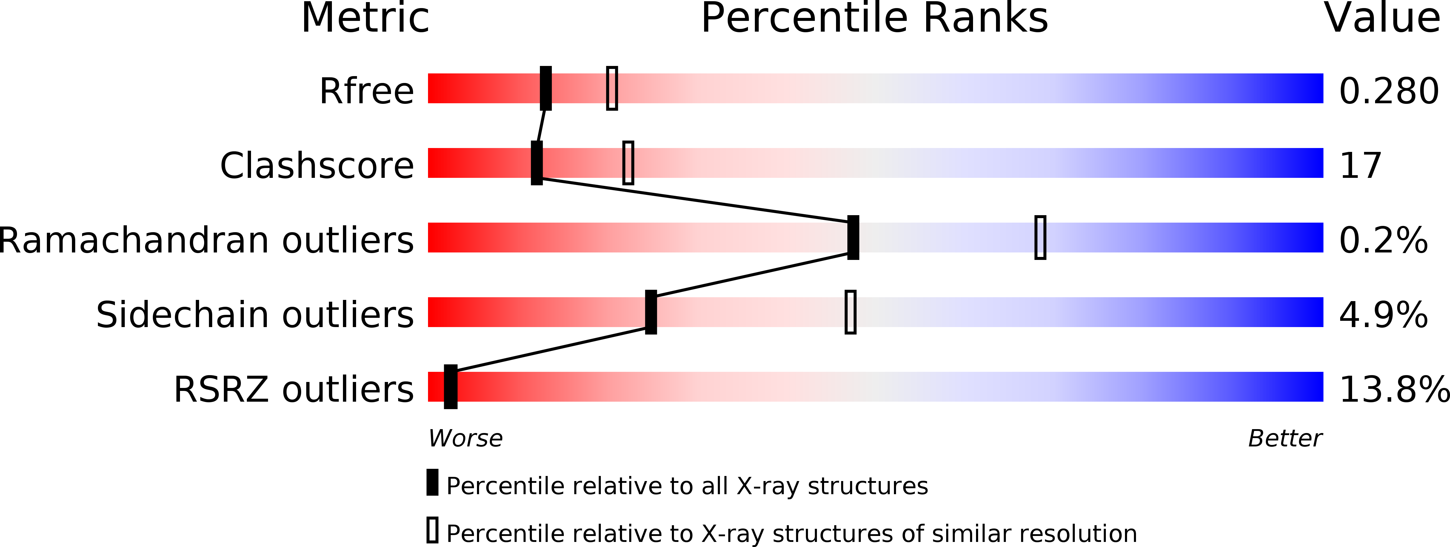
Resolution:
2.50 Å
R-Value Free:
0.29
R-Value Work:
0.23
Space Group:
P 21 21 21
Macromolecular Entities
Structures with similar UniProt ID
Protein Blast
Polymer Type:polypeptide(L)
Molecule:Cathepsin L1
Gene (Uniprot):CTSL
Chain IDs:A, B
Chain Length:220
Number of Molecules:2
Biological Source:Homo sapiens
Modified Residue
Compound ID Chain ID Parent Comp ID Details 2D Image
CSD A CYS 3-SULFINOALANINE
Ligand Molecules
Primary Citation
A Combined Crystallographic and Molecular Dynamics Study of Cathepsin L Retrobinding Inhibitors
J.Med.Chem. ? ? ? (2009)
PMID: 19761244 DOI: 10.1021/jm900596y

Abstact

We report the crystal structures of three noncovalent retrobinding inhibitors in complex with mature cathepsin L up to resolutions of 2.5, 1.8, and 2.5 A, respectively. These inhibitors were Bpa-(Nepsilon-Bpa)Lys-DArg-Tyr-Npe, Bpa-(Nepsilon-Bpa)Lys-DArg-Phe-Npe, and Bpa-MCys-DArg-Phe-Npe, where Bpa = biphenylacetyl and Pea = N-phenylethyl. These were selected to clarify the binding mode of the biphenyl groups in the S' subsites because the addition of a second biphenyl does not improve potency. Examination of the symmetry-related monomers in the crystal structures revealed inhibitor-inhibitor crystal packing interactions. Molecular dynamics simulations were then used to explore the structure and dynamical behavior of the isolated protein-ligand complexes in solution. In the simulations, the backbone biphenyl groups for all three inhibitors ended up in the same location despite having started out in different orientations in the initial crystal structure conformations. The lack of improved potency of the larger inhibitors over the smaller one is attributed to a correspondingly greater entropic cost of binding.

Legend

Protein

Chemical

Disease

Primary Citation of related structures