3H8A image
Deposition Date 2009-04-29
Release Date 2010-04-14
Last Version Date 2023-09-06
Entry Detail
PDB ID:
3H8A
Title:
Crystal structure of E. coli enolase bound to its cognate RNase E recognition domain
Biological Source:
Source Organism:
(Taxon ID: 83333)Escherichia coli (Taxon ID: 562)
Method Details:
Experimental Method:
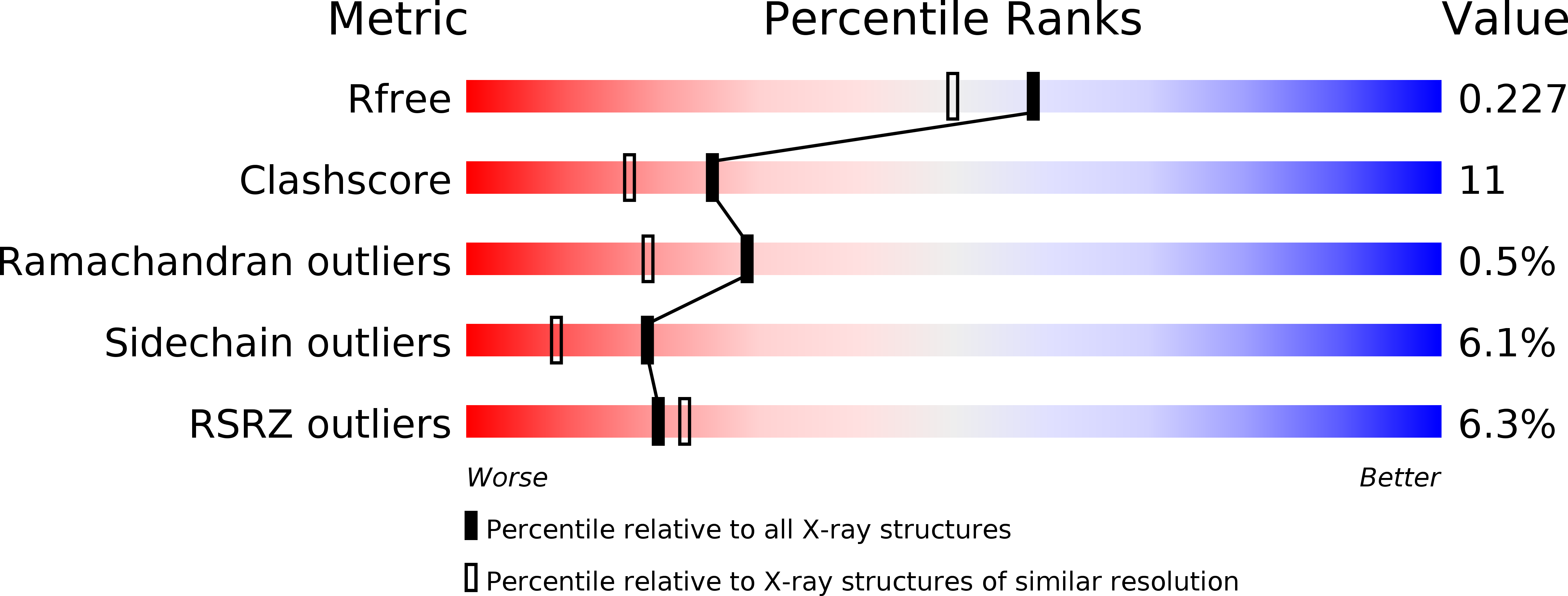
Resolution:
1.90 Å
R-Value Free:
0.22
R-Value Work:
0.18
R-Value Observed:
0.18
Space Group:
P 21 21 21
Macromolecular Entities
Polymer Type:polypeptide(L)
Molecule:Enolase
Gene (Uniprot):eno
Chain IDs:A, B, C, D
Chain Length:432
Number of Molecules:4
Biological Source:Escherichia coli
Polymer Type:polypeptide(L)
Molecule:RNase E
Chain IDs:E, F
Chain Length:28
Number of Molecules:2
Biological Source:
Ligand Molecules
Primary Citation
Molecular recognition between Escherichia coli enolase and ribonuclease E.
Acta Crystallogr.,Sect.D 66 1036 1040 (2010)
PMID: 20823555 DOI: 10.1107/S0907444910030015

Abstact

In Escherichia coli and many other bacterial species, the glycolytic enzyme enolase is a component of the multi-enzyme RNA degradosome, an assembly that is involved in RNA processing and degradation. Enolase is recruited into the degradosome through interactions with a small recognition motif located within the degradosome-scaffolding domain of RNase E. Here, the crystal structure of enolase bound to its cognate site from RNase E (residues 823-850) at 1.9 A resolution is presented. The structure suggests that enolase may help to organize an adjacent conserved RNA-binding motif in RNase E.

Legend

Protein

Chemical

Disease

Primary Citation of related structures