Deposition Date
2009-04-24
Release Date
2010-04-28
Last Version Date
2024-11-06
Entry Detail
PDB ID:
3H7D
Keywords:
Title:
The crystal structure of the cathepsin K Variant M5 in complex with chondroitin-4-sulfate
Biological Source:
Source Organism(s):
Homo sapiens (Taxon ID: 9606)
Expression System(s):
Method Details:
Experimental Method:
Resolution:
2.24 Å
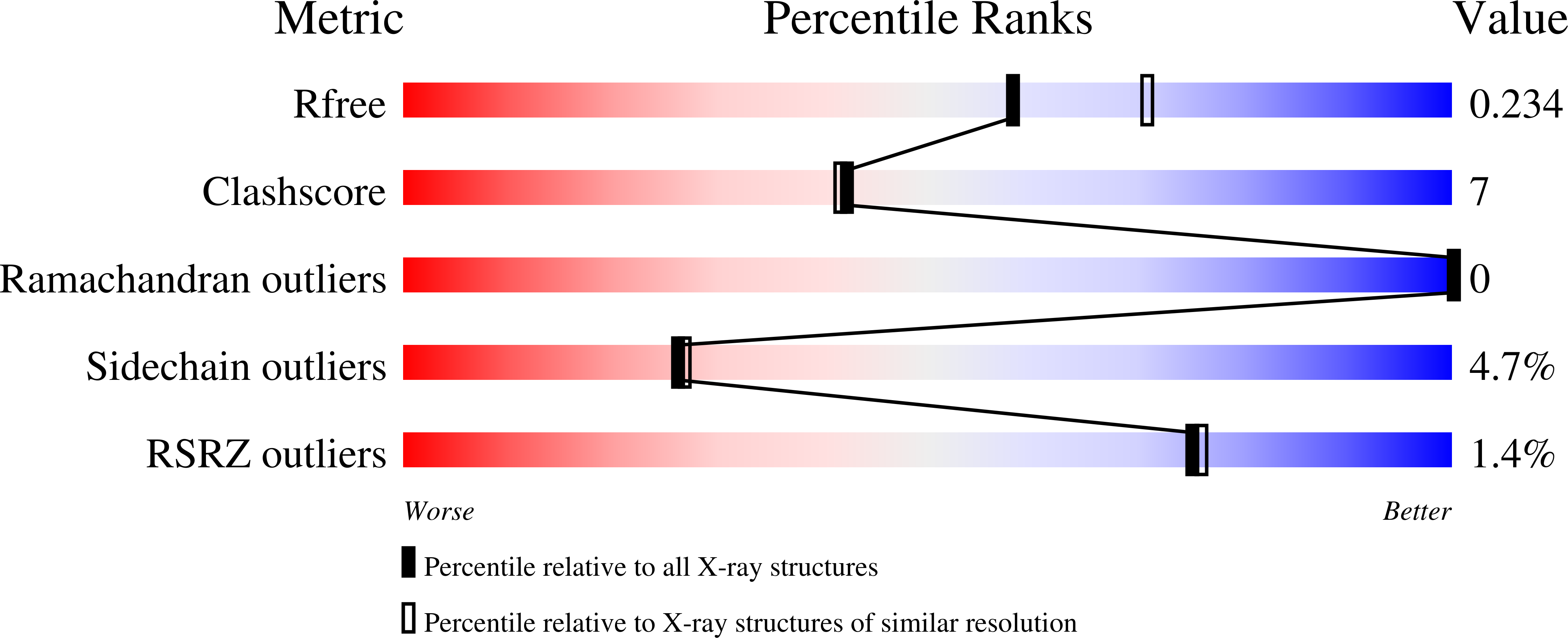
R-Value Free:
0.23
R-Value Work:
0.17
R-Value Observed:
0.18
Space Group:
C 1 2 1