Deposition Date
2009-04-24
Release Date
2009-05-12
Last Version Date
2024-11-06
Entry Detail
PDB ID:
3H75
Keywords:
Title:
Crystal Structure of a Periplasmic Sugar-binding protein from the Pseudomonas fluorescens
Biological Source:
Source Organism(s):
Pseudomonas fluorescens Pf-5 (Taxon ID: 220664)
Expression System(s):
Method Details:
Experimental Method:
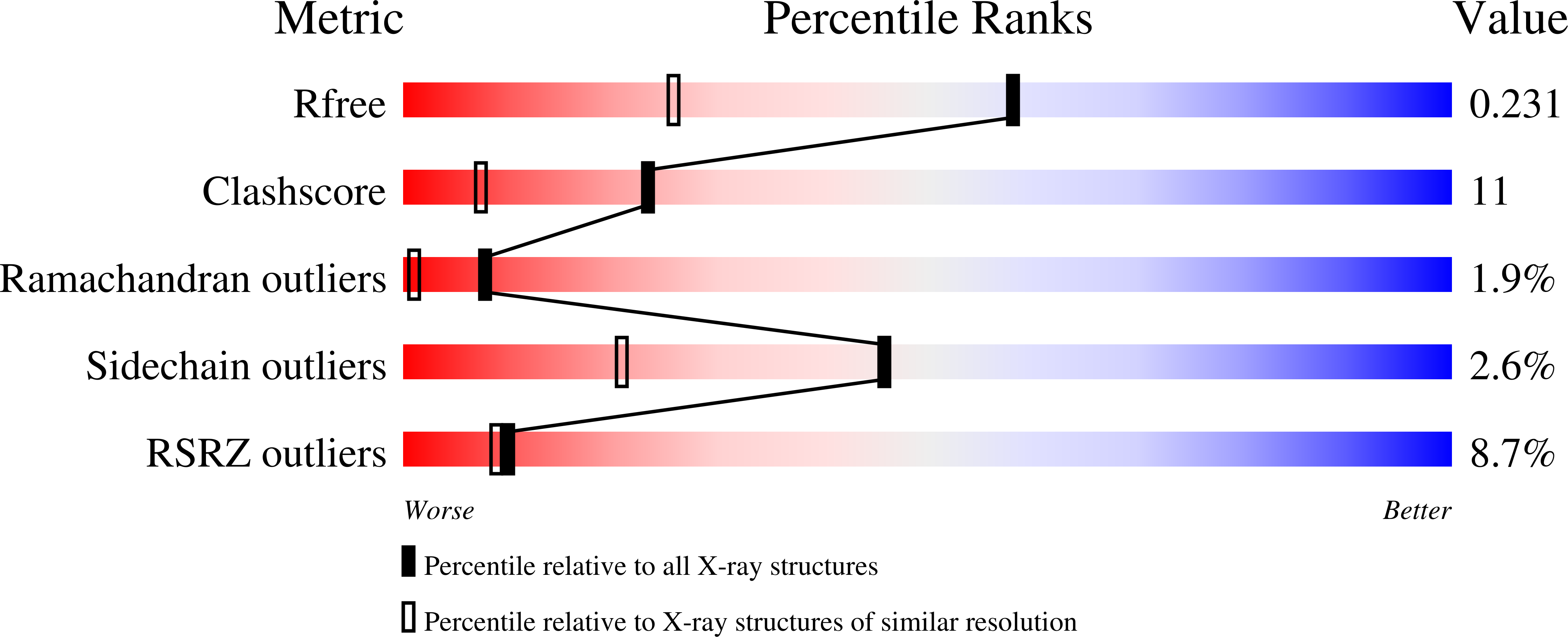
Resolution:
1.60 Å
R-Value Free:
0.23
R-Value Work:
0.20
R-Value Observed:
0.20
Space Group:
P 21 21 2