3H5C image
Deposition Date 2009-04-21
Release Date 2010-04-28
Last Version Date 2024-11-27
Entry Detail
PDB ID:
3H5C
Title:
X-Ray Structure of Protein Z-Protein Z Inhibitor Complex
Biological Source:
Source Organism:
Homo sapiens (Taxon ID: 9606)
Host Organism:
Method Details:
Experimental Method:
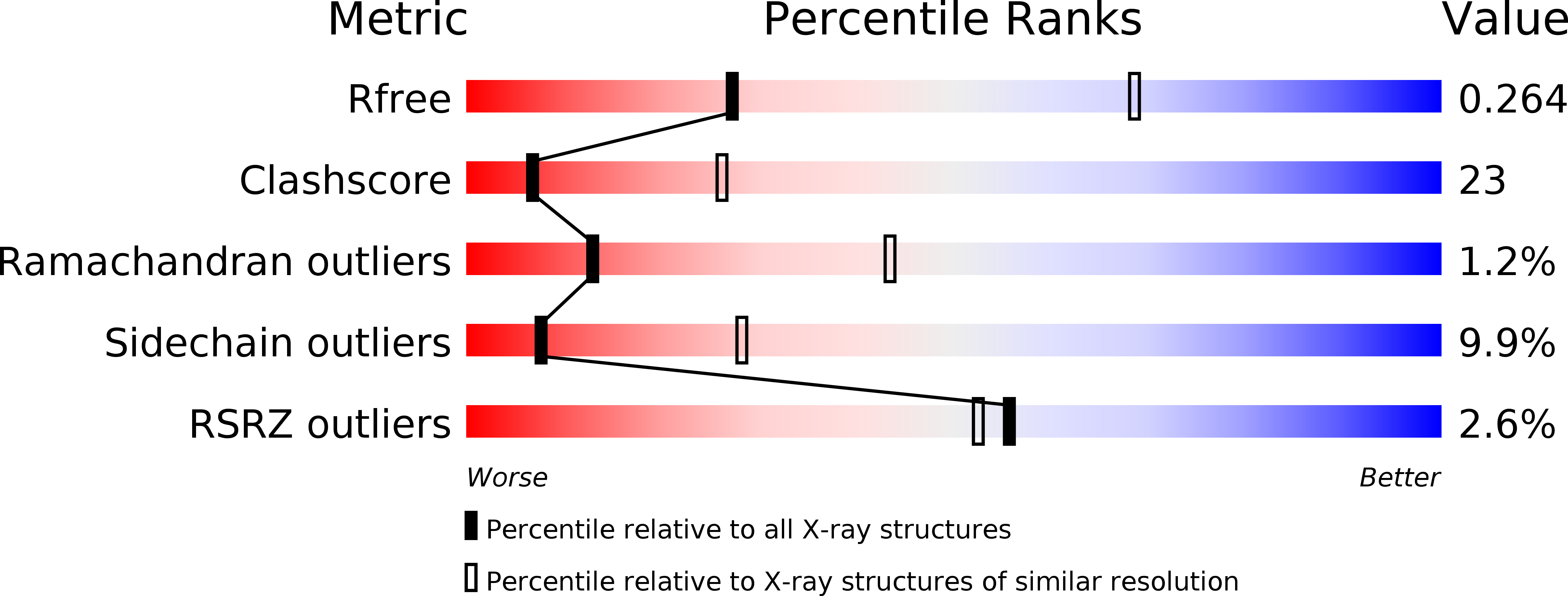
Resolution:
3.26 Å
R-Value Free:
0.26
R-Value Work:
0.20
R-Value Observed:
0.20
Space Group:
P 65 2 2
Macromolecular Entities
Polymer Type:polypeptide(L)
Molecule:Protein Z-dependent protease inhibitor
Gene (Uniprot):SERPINA10
Chain IDs:A
Chain Length:423
Number of Molecules:1
Biological Source:Homo sapiens
Polymer Type:polypeptide(L)
Molecule:Vitamin K-dependent protein Z
Gene (Uniprot):PROZ
Chain IDs:B
Chain Length:317
Number of Molecules:1
Biological Source:Homo sapiens
Modified Residue
Compound ID Chain ID Parent Comp ID Details 2D Image
ASN A ASN GLYCOSYLATION SITE
Primary Citation
Basis for the specificity and activation of the serpin protein Z-dependent proteinase inhibitor (ZPI) as an inhibitor of membrane-associated factor Xa.
J.Biol.Chem. 285 20399 20409 (2010)
PMID: 20427285 DOI: 10.1074/jbc.M110.112748

Abstact

The serpin ZPI is a protein Z (PZ)-dependent specific inhibitor of membrane-associated factor Xa (fXa) despite having an unfavorable P1 Tyr. PZ accelerates the inhibition reaction approximately 2000-fold in the presence of phospholipid and Ca(2+). To elucidate the role of PZ, we determined the x-ray structure of Gla-domainless PZ (PZ(DeltaGD)) complexed with protein Z-dependent proteinase inhibitor (ZPI). The PZ pseudocatalytic domain bound ZPI at a novel site through ionic and polar interactions. Mutation of four ZPI contact residues eliminated PZ binding and membrane-dependent PZ acceleration of fXa inhibition. Modeling of the ternary Michaelis complex implicated ZPI residues Glu-313 and Glu-383 in fXa binding. Mutagenesis established that only Glu-313 is important, contributing approximately 5-10-fold to rate acceleration of fXa and fXIa inhibition. Limited conformational change in ZPI resulted from PZ binding, which contributed only approximately 2-fold to rate enhancement. Instead, template bridging from membrane association, together with previously demonstrated interaction of the fXa and ZPI Gla domains, resulted in an additional approximately 1000-fold rate enhancement. To understand why ZPI has P1 tyrosine, we examined a P1 Arg variant. This reacted at a diffusion-limited rate with fXa, even without PZ, and predominantly as substrate, reflecting both rapid acylation and deacylation. P1 tyrosine thus ensures that reaction with fXa or most other arginine-specific proteinases is insignificant unless PZ binds and localizes ZPI and fXa on the membrane, where the combined effects of Gla-Gla interaction, template bridging, and interaction of fXa with Glu-313 overcome the unfavorability of P1 Tyr and ensure a high rate of reaction as an inhibitor.

Legend

Protein

Chemical

Disease

Primary Citation of related structures