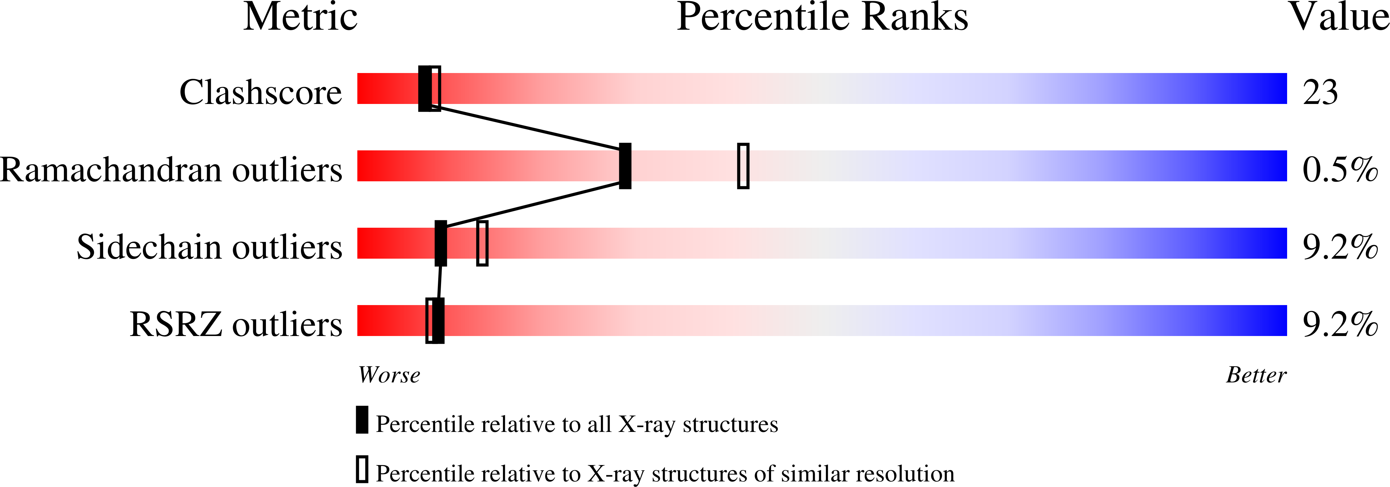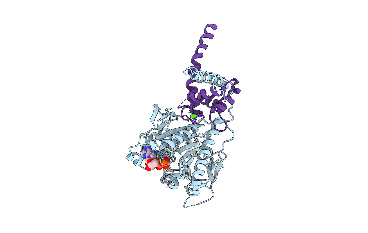
3H4S image
Deposition Date 2009-04-20
Release Date 2009-05-19
Last Version Date 2023-09-06
Entry Detail
PDB ID:
3H4S
Title:
Structure of the complex of a mitotic kinesin with its calcium binding regulator
Biological Source:
Source Organism:
Host Organism:
Method Details:
Experimental Method:
Resolution:
2.40 Å
R-Value Free:
0.22
R-Value Work:
0.22
R-Value Observed:
0.22
Space Group:
P 65 2 2
Macromolecular Entities
Polymer Type:polypeptide(L)
Molecule:Kinesin-like calmodulin-binding protein
Gene (Uniprot):KIN14E
Mutations:C1131N
Chain IDs:A
Chain Length:386
Number of Molecules:1
Biological Source:Arabidopsis thaliana
Polymer Type:polypeptide(L)
Molecule:KCBP interacting Ca2+-binding protein
Gene (Uniprot):KIC
Chain IDs:B (auth: E)
Chain Length:135
Number of Molecules:1
Biological Source:Arabidopsis thaliana
Primary Citation
Structure of the complex of a mitotic kinesin with its calcium binding regulator.
Proc.Natl.Acad.Sci.USA 106 8175 8179 (2009)
PMID: 19416847 DOI: 10.1073/pnas.0811131106

Abstact

Much of the transport, tension, and movement in mitosis depends on kinesins, the ATP-powered microtubule-based motors. We report the crystal structure of a kinesin complex, the mitotic kinesin KCBP bound to its principal regulator KIC. Shown to be a Ca(2+) sensor, KIC works as an allosteric trap. Extensive intermolecular interactions with KIC stabilize kinesin in its ADP-bound conformation. A critical component of the kinesin motile mechanism, called the neck mimic, switches its association from kinesin to KIC, stalling the motor. KIC denies access of the motor to its track by steric interference. Two major features of this regulation, allosteric trapping and steric blocking, are likely to be general for all kinesins.

Legend

Protein

Chemical

Disease

Primary Citation of related structures