3H4R image
Deposition Date 2009-04-20
Release Date 2009-05-26
Last Version Date 2024-02-21
Entry Detail
PDB ID:
3H4R
Keywords:
Title:
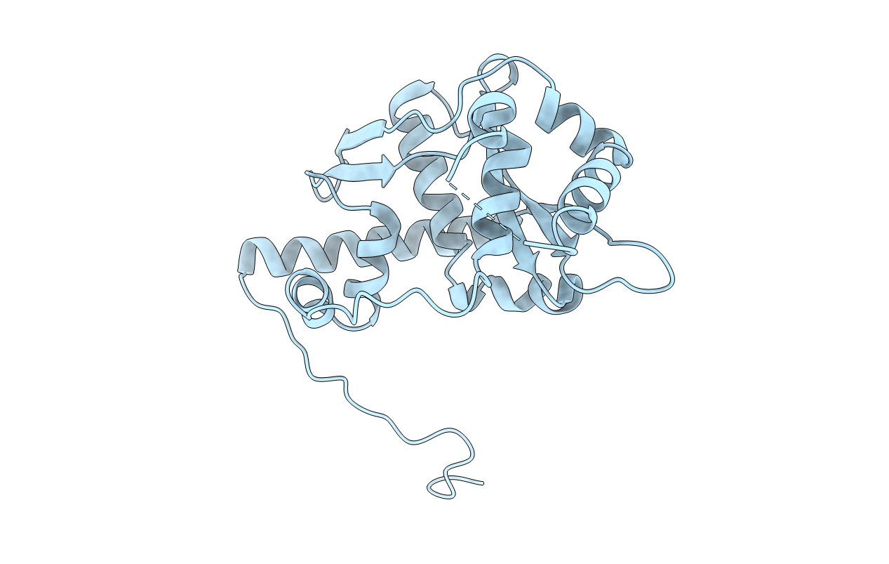
Crystal structure of E. coli RecE exonuclease
Biological Source:
Source Organism:
Host Organism:
Method Details:
Experimental Method:
Resolution:
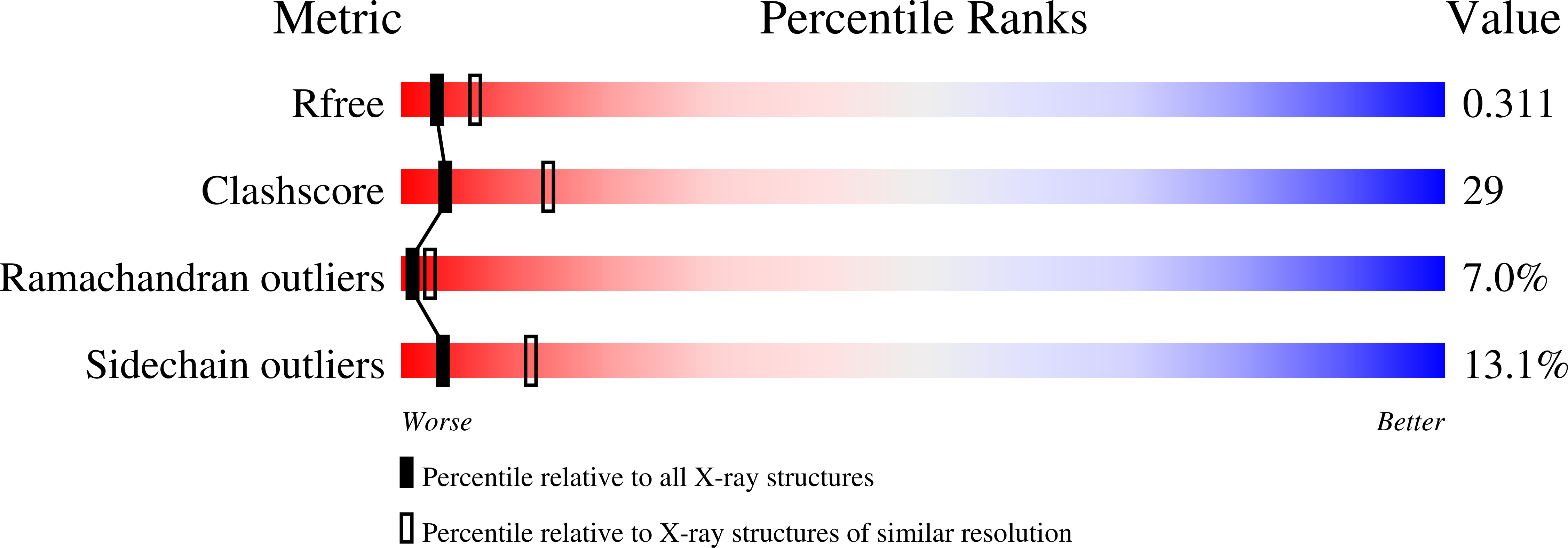
2.80 Å
R-Value Free:
0.31
R-Value Work:
0.29
R-Value Observed:
0.29
Space Group:
P 4 21 2
Macromolecular Entities
Polymer Type:polypeptide(L)
Molecule:Exodeoxyribonuclease 8
Gene (Uniprot):recE
Mutations:P658L
Chain IDs:A
Chain Length:265
Number of Molecules:1
Biological Source:Escherichia coli
Ligand Molecules
Primary Citation
Crystal structure of E. coli RecE protein reveals a toroidal tetramer for processing double-stranded DNA breaks.
Structure 17 690 702 (2009)
PMID: 19446525 DOI: 10.1016/j.str.2009.03.008

Abstact

Escherichia coli RecE protein is part of the classical RecET recombination system that has recently been used in powerful new methods for genetic engineering. RecE binds to free double-stranded DNA (dsDNA) ends and processively digests the 5'-ended strand to form 5'-mononucleotides and a 3'-overhang that is a substrate for single strand annealing promoted by RecT. Here, we report the crystal structure of the C-terminal nuclease domain of RecE at 2.8 A resolution. RecE forms a toroidal tetramer with a central tapered channel that is wide enough to bind dsDNA at one end, but is partially plugged at the other end by the C-terminal segment of the protein. Four narrow tunnels, one within each subunit of the tetramer, lead from the central channel to the four active sites, which lie about 15 A from the channel. The structure, combined with mutational studies, suggests a mechanism in which dsDNA enters through the open end of the central channel, the 5'-ended strand passes through a tunnel to access one of the four active sites, and the 3'-ended strand passes through the plugged end of the channel at the back of the tetramer.

Legend

Protein

Chemical

Disease

Primary Citation of related structures