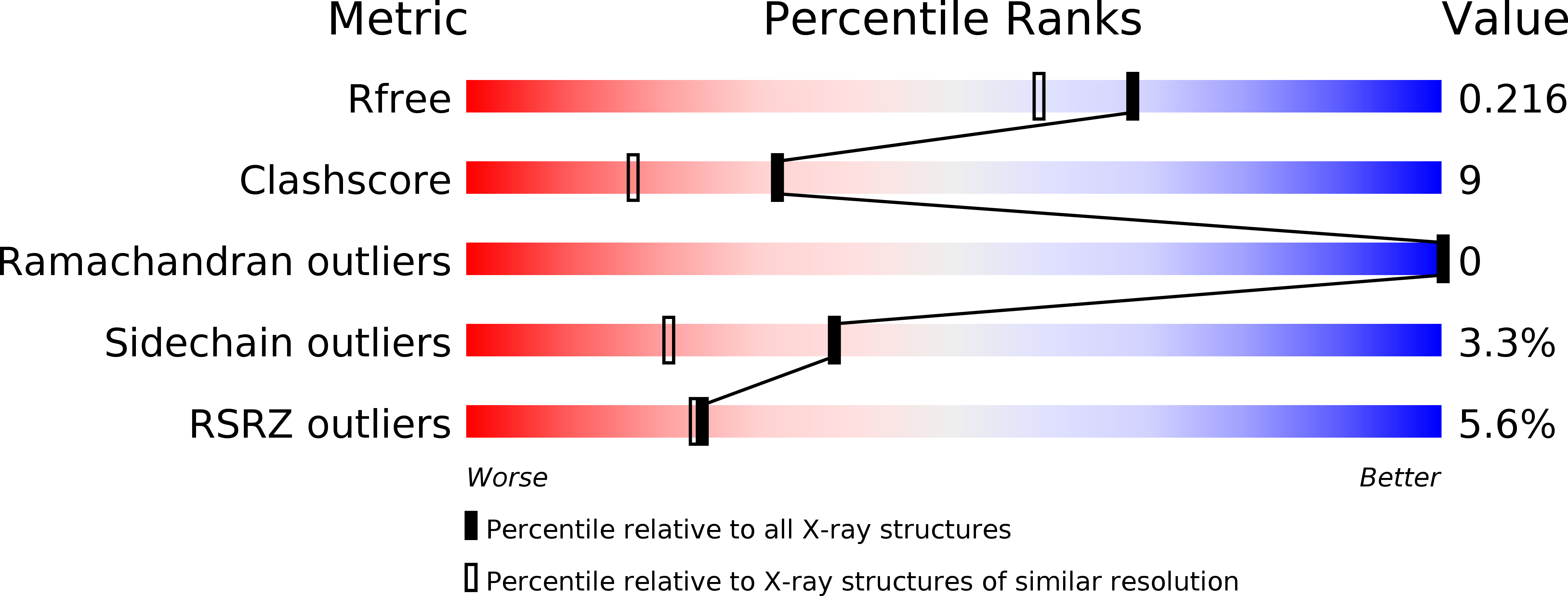
3H4G image
Deposition Date 2009-04-20
Release Date 2009-05-05
Last Version Date 2023-11-01
Entry Detail
PDB ID:
3H4G
Keywords:
Title:
Structure of aldehyde reductase holoenzyme in complex with potent aldose reductase inhibitor Fidarestat: Implications for inhibitor binding and selectivity
Biological Source:
Source Organism:
Sus scrofa (Taxon ID: 9823)
Method Details:
Experimental Method:
Resolution:
1.85 Å
R-Value Free:
0.21
R-Value Work:
0.18
R-Value Observed:
0.18
Space Group:
P 65 2 2
Macromolecular Entities
Polymer Type:polypeptide(L)
Molecule:Alcohol dehydrogenase [NADP+]
Gene (Uniprot):AKR1A1
Chain IDs:A
Chain Length:325
Number of Molecules:1
Biological Source:Sus scrofa
Primary Citation
Structure of aldehyde reductase holoenzyme in complex with the potent aldose reductase inhibitor fidarestat: implications for inhibitor binding and selectivity
J.Med.Chem. 48 5536 5542 (2005)
PMID: 16107153 DOI: 10.1021/jm050412o

Abstact

Structure determination of porcine aldehyde reductase holoenzyme in complex with the potent aldose reductase inhibitor fidarestat was carried out to explain the difference in the potency of the inhibitor for aldose and aldehyde reductases. The hydrogen bonds between the active-site residues Tyr50, His113, and Trp114 and fidarestat are conserved in the two enzymes. In aldose reductase, Leu300 forms a hydrogen bond through its main-chain nitrogen atom with the exocyclic amide group of the inhibitor, which when replaced with a Pro in aldehyde reductase, cannot form a hydrogen bond, thus causing a loss in binding energy. Furthermore, in aldehyde reductase, the side chain of Trp220 occupies a disordered split conformation that is not observed in aldose reductase. Molecular modeling and inhibitory activity measurements suggest that the difference in the interaction between the side chain of Trp220 and fidarestat may contribute to the difference in the binding of the inhibitor to the enzymes.

Legend

Protein

Chemical

Disease

Primary Citation of related structures