3H3U image
Deposition Date 2009-04-17
Release Date 2010-02-02
Last Version Date 2023-11-01
Entry Detail
PDB ID:
3H3U
Keywords:
Title:
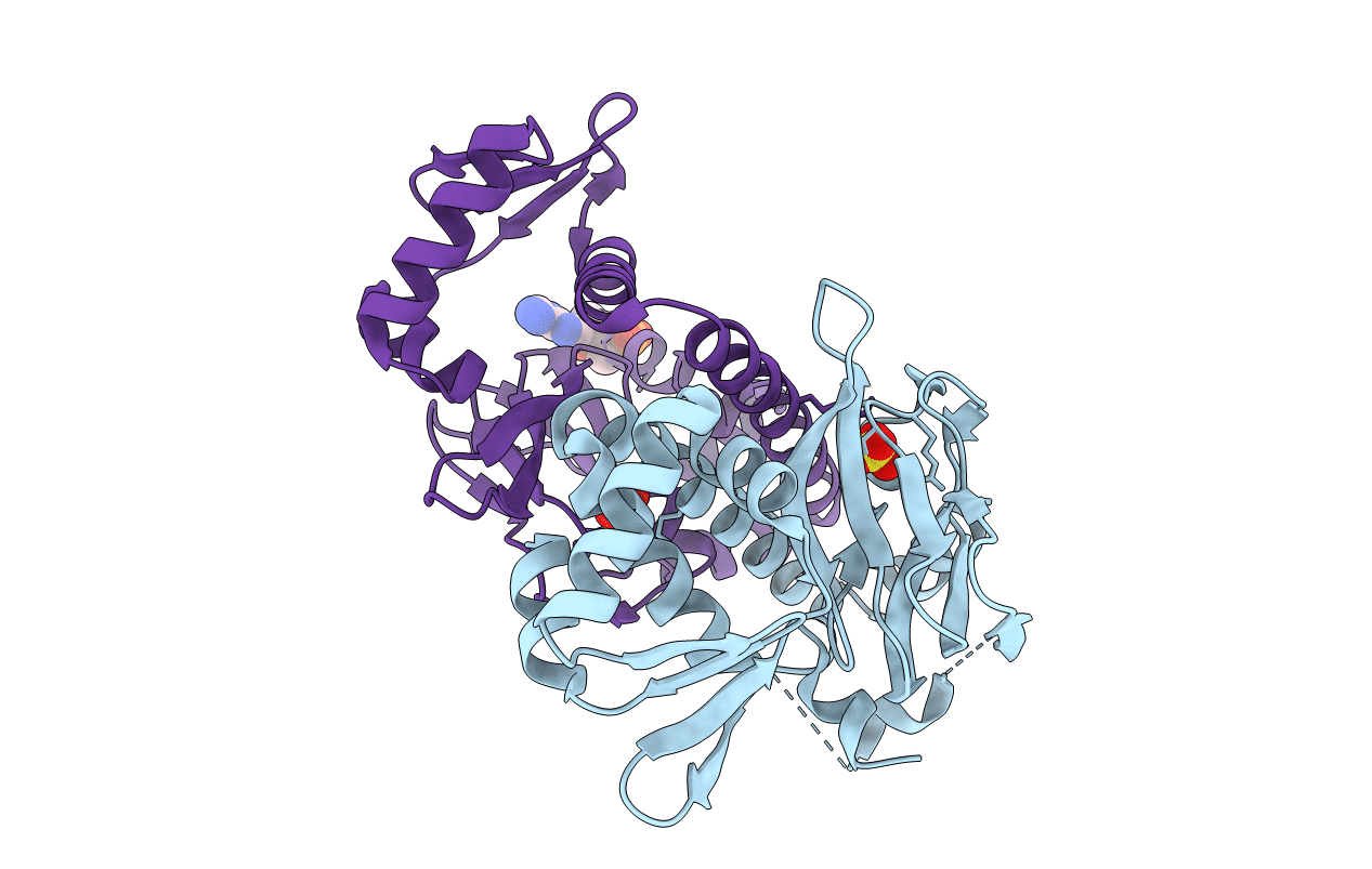
Crystal structure of CRP (cAMP receptor Protein) from Mycobacterium tuberculosis
Biological Source:
Source Organism:
Host Organism:
Method Details:
Experimental Method:
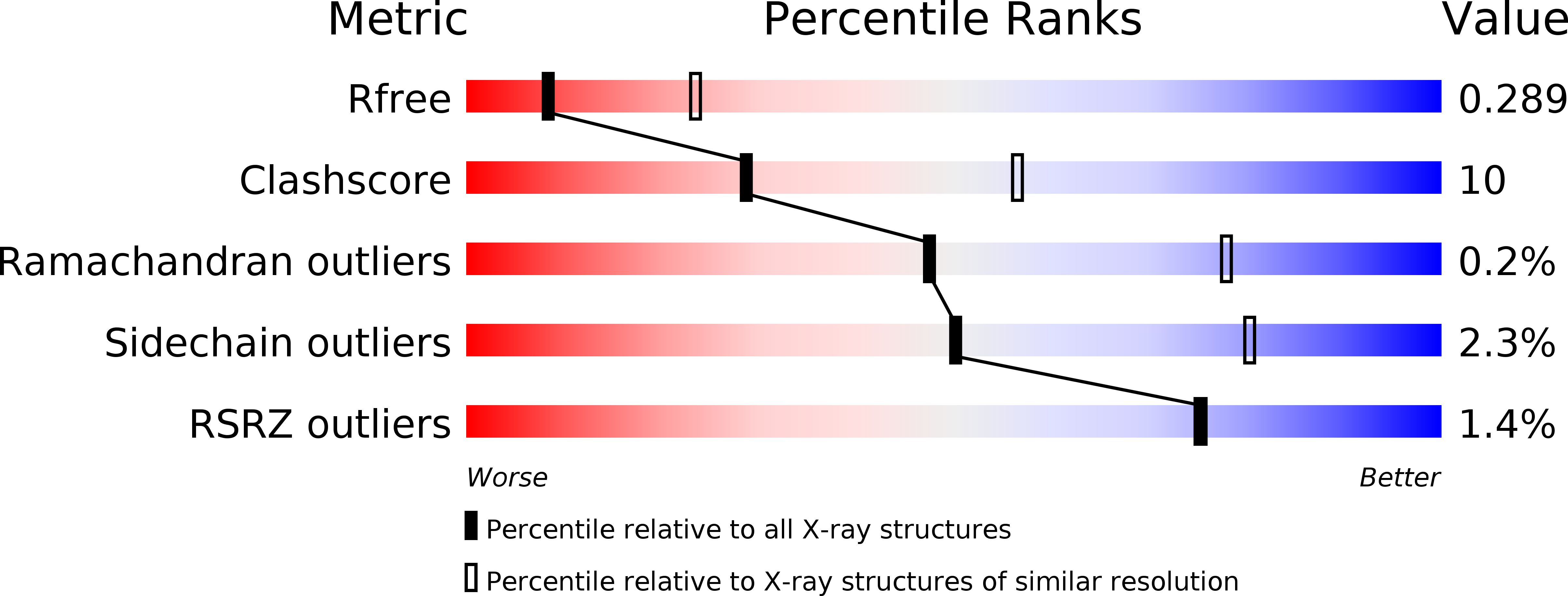
Resolution:
2.90 Å
R-Value Free:
0.29
R-Value Work:
0.22
R-Value Observed:
0.22
Space Group:
P 21 21 21
Macromolecular Entities
Polymer Type:polypeptide(L)
Molecule:PROBABLE TRANSCRIPTIONAL REGULATORY PROTEIN (PROBABLY CRP/FNR-FAMILY)
Chain IDs:A, B
Chain Length:224
Number of Molecules:2
Biological Source:Mycobacterium tuberculosis
Primary Citation
Mapping conformational transitions in cyclic AMP receptor protein: crystal structure and normal-mode analysis of Mycobacterium tuberculosis apo-cAMP receptor protein
Biophys.J. 98 305 314 (2010)
PMID: 20338852 DOI: 10.1016/j.bpj.2009.10.016

Abstact

Cyclic AMP (cAMP) receptor protein, which acts as the sensor of cAMP levels in cells, is a well-studied transcription factor that is best known for allosteric changes effected by the binding of cAMP. Although genetic and biochemical data on the protein are available from several sources, structural information about the cAMP-free protein has been lacking. Therefore, the precise atomic events that take place upon binding of cAMP, leading to conformational changes in the protein and its activation to bind DNA, have been elusive. In this work we solved the cAMP-free crystal structure of the Mycobacterium tuberculosis homolog of cAMP receptor protein at 2.9 A resolution, and carried out normal-mode analysis to map conformational transitions among its various conformational states. In our structure, the cAMP-binding domain holds onto the DNA-binding domain via strong hydrophobic interactions, thereby freezing the latter in a conformation that is not competent to bind DNA. The two domains release each other in the presence of cAMP, making the DNA-binding domain more flexible and allowing it to bind its cognate DNA via an induced-fit mechanism. The structure of the cAMP-free protein and results of the normal-mode analysis therefore highlight an elegant mechanism of the allosteric changes effected by the binding of cAMP.

Legend

Protein

Chemical

Disease

Primary Citation of related structures