Deposition Date
2009-04-16
Release Date
2009-08-11
Last Version Date
2024-11-27
Entry Detail
PDB ID:
3H3G
Keywords:
Title:
Crystal structure of the extracellular domain of the human parathyroid hormone receptor (PTH1R) in complex with parathyroid hormone-related protein (PTHrP)
Biological Source:
Source Organism(s):
Escherichia coli (Taxon ID: 562)
Homo sapiens (Taxon ID: 9606)
Homo sapiens (Taxon ID: 9606)
Expression System(s):
Method Details:
Experimental Method:
Resolution:
1.94 Å
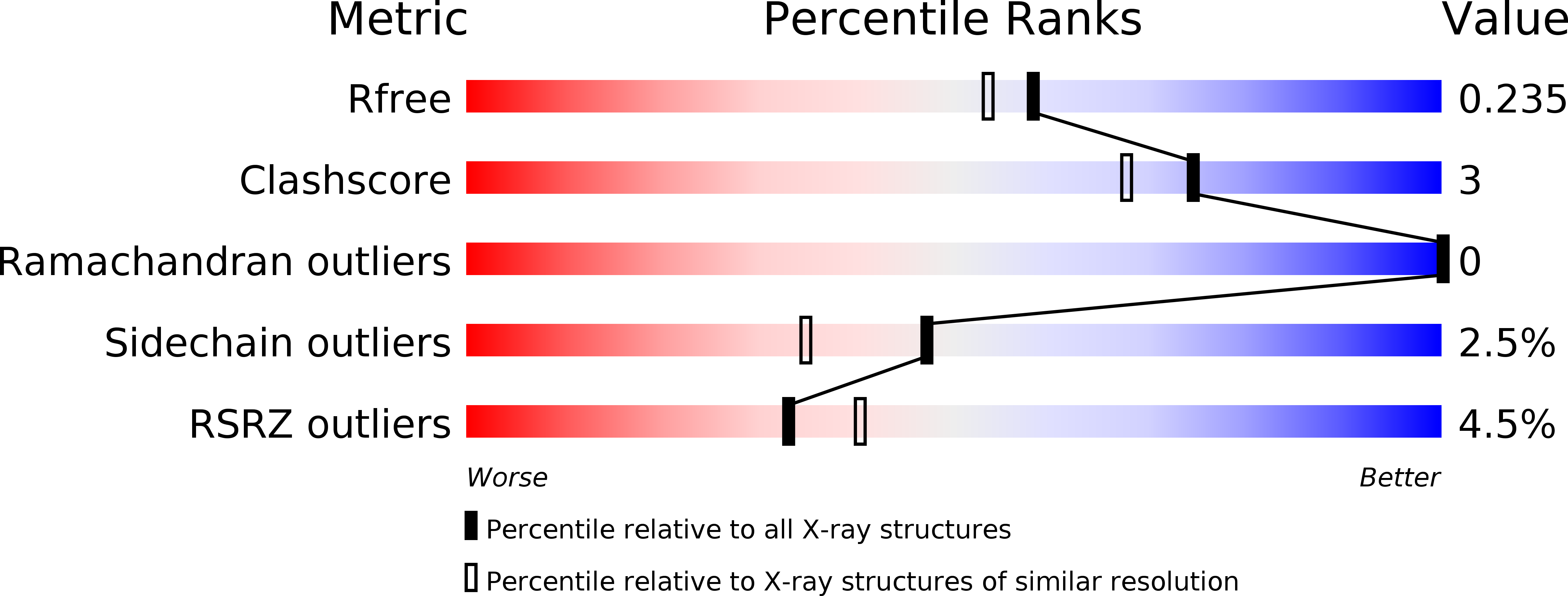
R-Value Free:
0.23
R-Value Work:
0.19
R-Value Observed:
0.19
Space Group:
P 41 21 2