Deposition Date
2009-04-13
Release Date
2009-09-08
Last Version Date
2025-03-26
Entry Detail
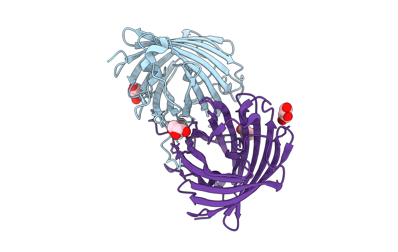
Biological Source:
Source Organism(s):
Entacmaea Quadricolor (Taxon ID: 6118)
Expression System(s):
Method Details:
Experimental Method:
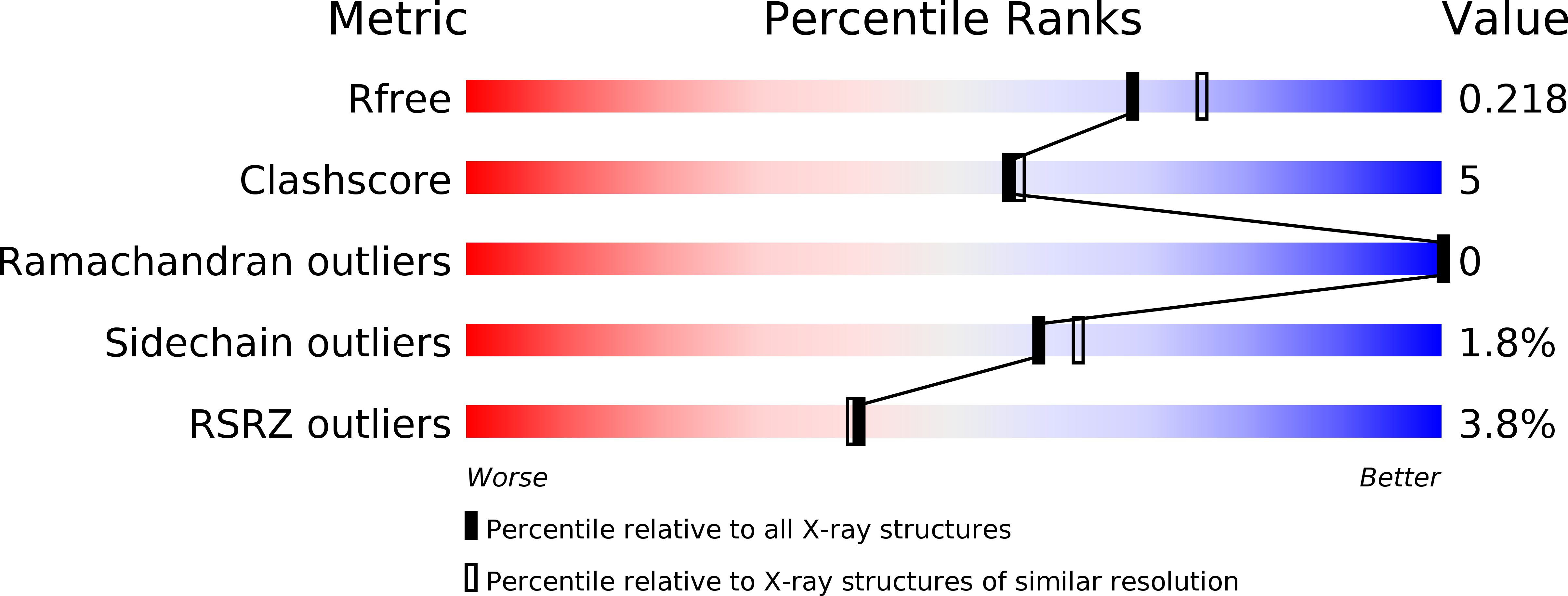
Resolution:
2.00 Å
R-Value Free:
0.22
R-Value Work:
0.17
Space Group:
I 4