3H1C image
Deposition Date 2009-04-11
Release Date 2009-05-19
Last Version Date 2023-11-01
Entry Detail
PDB ID:
3H1C
Keywords:
Title:
Crystal structure of Polynucleotide Phosphorylase (PNPase) core bound to RNase E and Tungstate
Biological Source:
Source Organism:
(Taxon ID: )Escherichia coli (Taxon ID: 562)
Method Details:
Experimental Method:
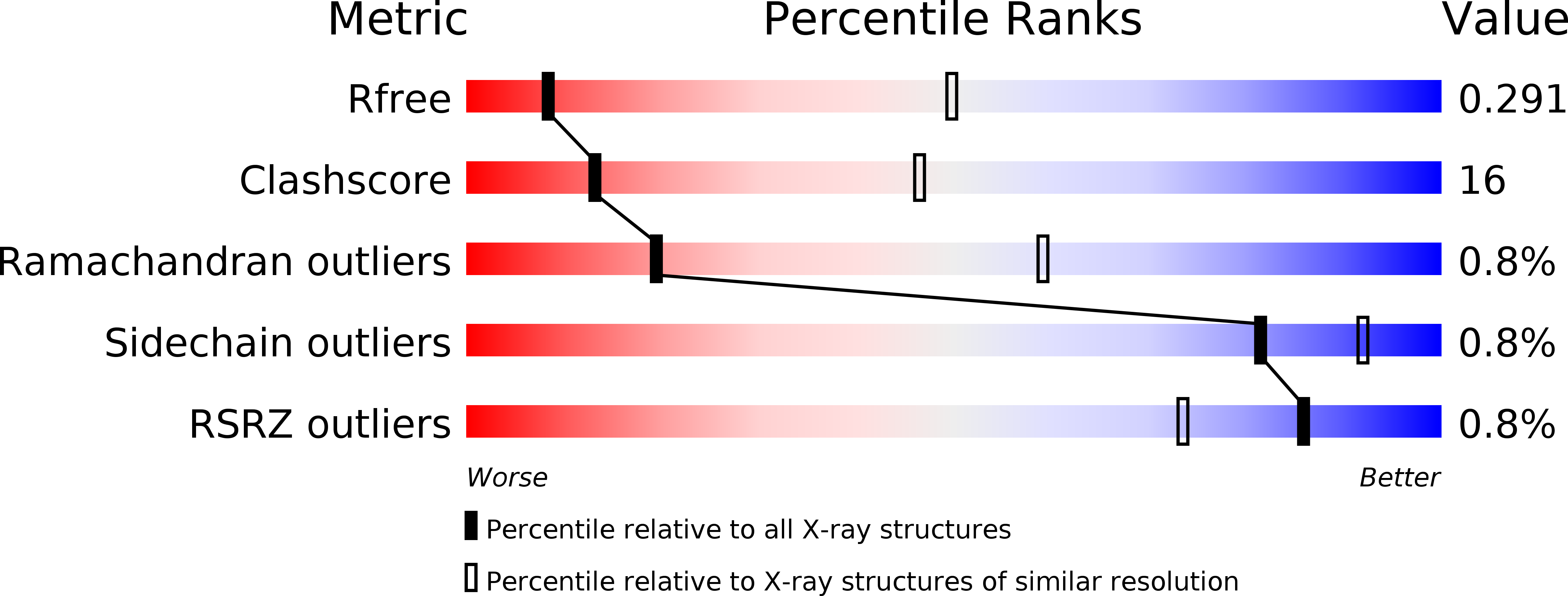
Resolution:
3.57 Å
R-Value Free:
0.30
R-Value Work:
0.27
R-Value Observed:
0.30
Space Group:
P 21 21 21
Macromolecular Entities
Polymer Type:polypeptide(L)
Molecule:Polyribonucleotide nucleotidyltransferase
Gene (Uniprot):pnp
Chain IDs:A, C (auth: B), E (auth: C), G, I, K, M, O, Q (auth: R), S (auth: T), U (auth: V), W (auth: X)
Chain Length:549
Number of Molecules:12
Biological Source:Escherichia coli
Polymer Type:polypeptide(L)
Molecule:Ribonuclease E
Gene (Uniprot):rne
Chain IDs:B (auth: D), D (auth: E), F, H, J, L, N, P, R (auth: S), T (auth: U), V (auth: W), X (auth: Y)
Chain Length:41
Number of Molecules:12
Biological Source:
Ligand Molecules
Primary Citation
Crystal structure of Escherichia coli polynucleotide phosphorylase core bound to RNase E, RNA and manganese: implications for catalytic mechanism and RNA degradosome assembly
J.Mol.Biol. 389 17 33 (2009)
PMID: 19327365 DOI: 10.1016/j.jmb.2009.03.051

Abstact

Polynucleotide phosphorylase (PNPase) is a processive exoribonuclease that contributes to messenger RNA turnover and quality control of ribosomal RNA precursors in many bacterial species. In Escherichia coli, a proportion of the PNPase is recruited into a multi-enzyme assembly, known as the RNA degradosome, through an interaction with the scaffolding domain of the endoribonuclease RNase E. Here, we report crystal structures of E. coli PNPase complexed with the recognition site from RNase E and with manganese in the presence or in the absence of modified RNA. The homotrimeric PNPase engages RNase E on the periphery of its ring-like architecture through a pseudo-continuous anti-parallel beta-sheet. A similar interaction pattern occurs in the structurally homologous human exosome between the Rrp45 and Rrp46 subunits. At the centre of the PNPase ring is a tapered channel with an adjustable aperture where RNA bases stack on phenylalanine side chains and trigger structural changes that propagate to the active sites. Manganese can substitute for magnesium as an essential co-factor for PNPase catalysis, and our crystal structure of the enzyme in complex with manganese suggests how the metal is positioned to stabilise the transition state. We discuss the implications of these structural observations for the catalytic mechanism of PNPase, its processive mode of action, and its assembly into the RNA degradosome.

Legend

Protein

Chemical

Disease

Primary Citation of related structures