Deposition Date
2009-04-11
Release Date
2009-06-16
Last Version Date
2024-02-21
Entry Detail
PDB ID:
3H16
Keywords:
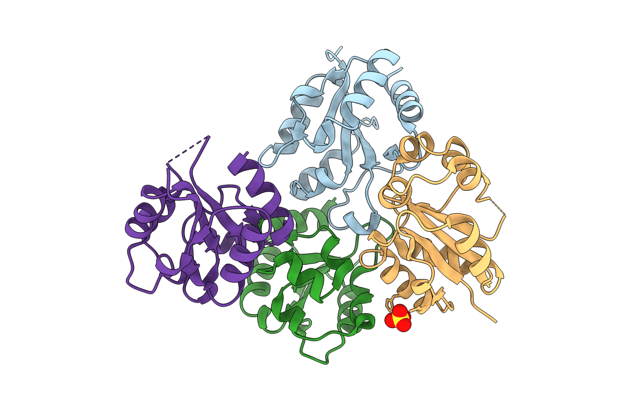
Title:
Crystal structure of a bacteria TIR domain, PdTIR from Paracoccus denitrificans
Biological Source:
Source Organism(s):
Paracoccus denitrificans PD1222 (Taxon ID: 318586)
Expression System(s):
Method Details:
Experimental Method:
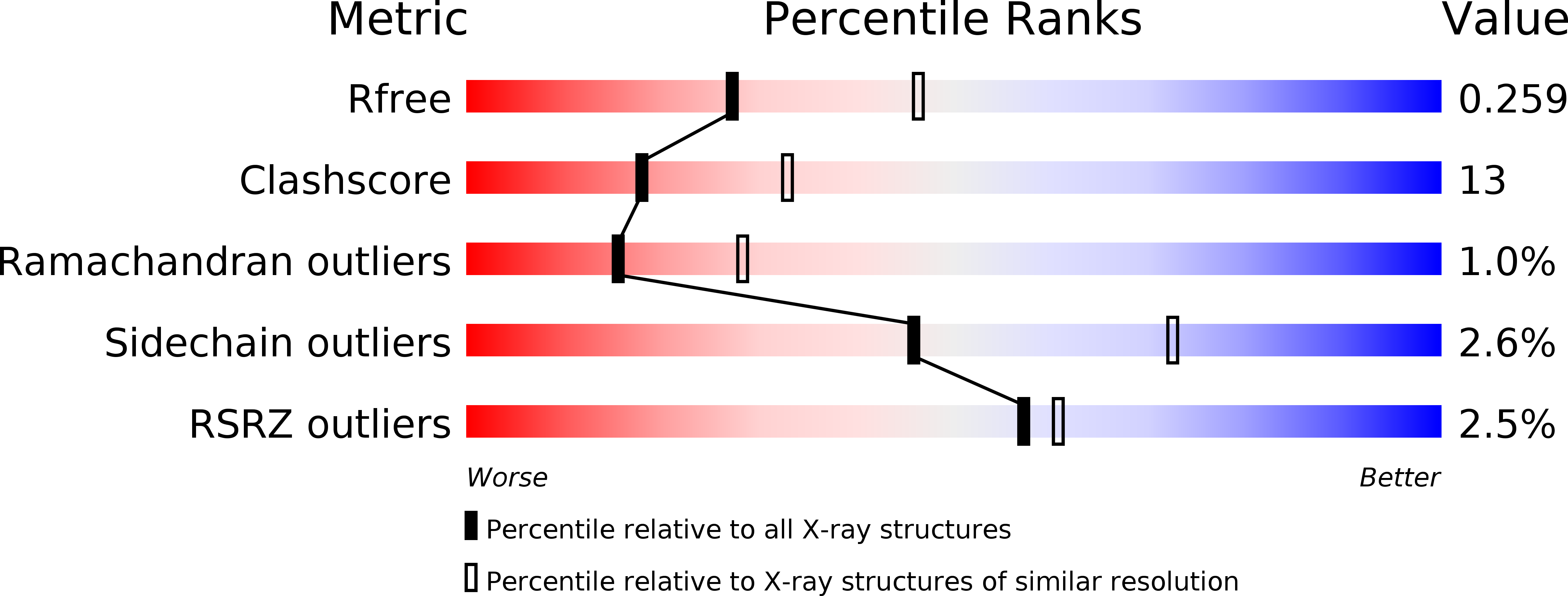
Resolution:
2.50 Å
R-Value Free:
0.24
R-Value Work:
0.19
R-Value Observed:
0.19
Space Group:
P 21 21 21