3GZX image
Deposition Date 2009-04-08
Release Date 2010-05-05
Last Version Date 2023-11-01
Entry Detail
PDB ID:
3GZX
Keywords:
Title:
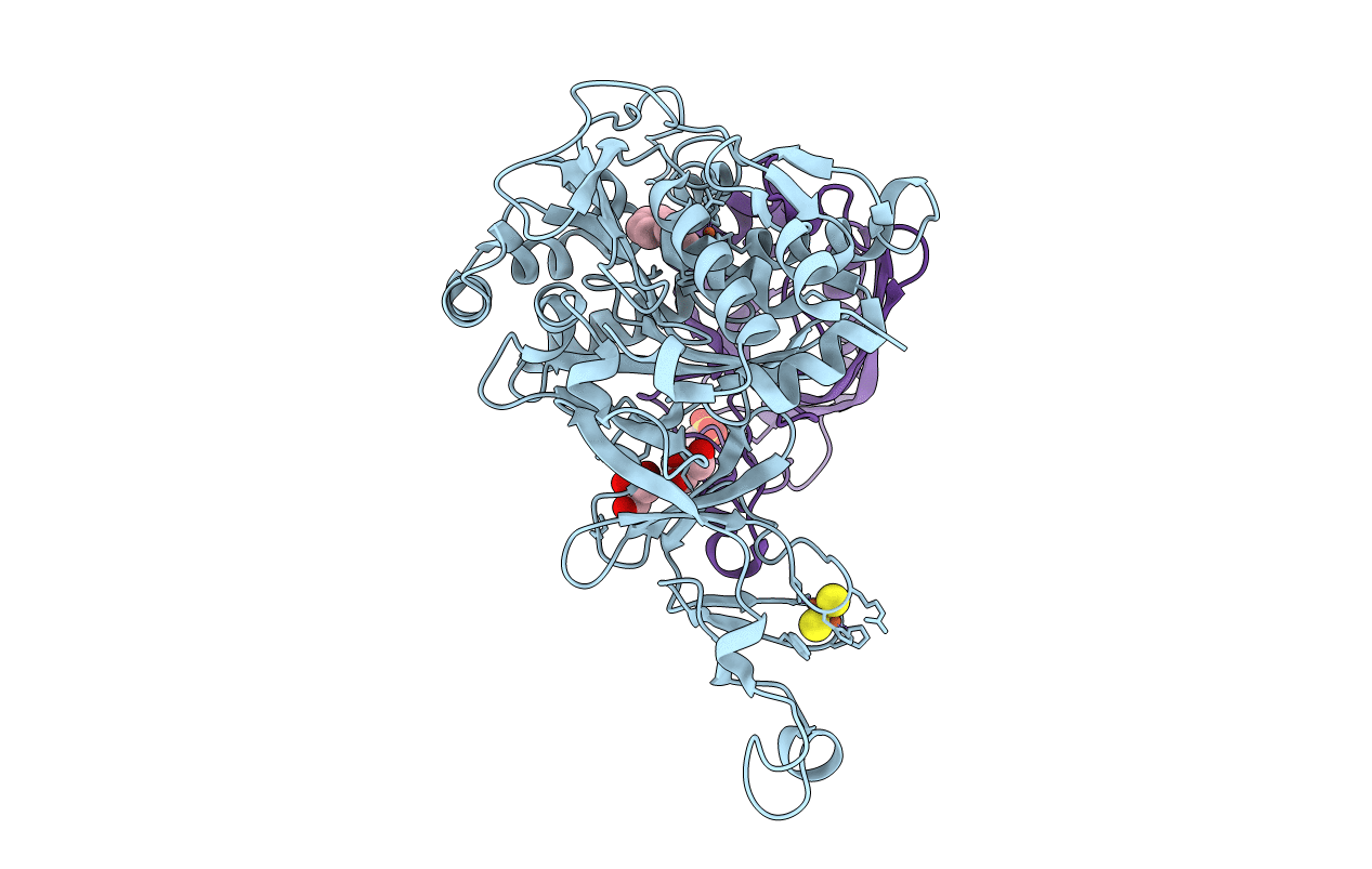
Crystal Structure of the Biphenyl Dioxygenase in complex with Biphenyl from Comamonas testosteroni Sp. Strain B-356
Biological Source:
Source Organism:
Host Organism:
Method Details:
Experimental Method:
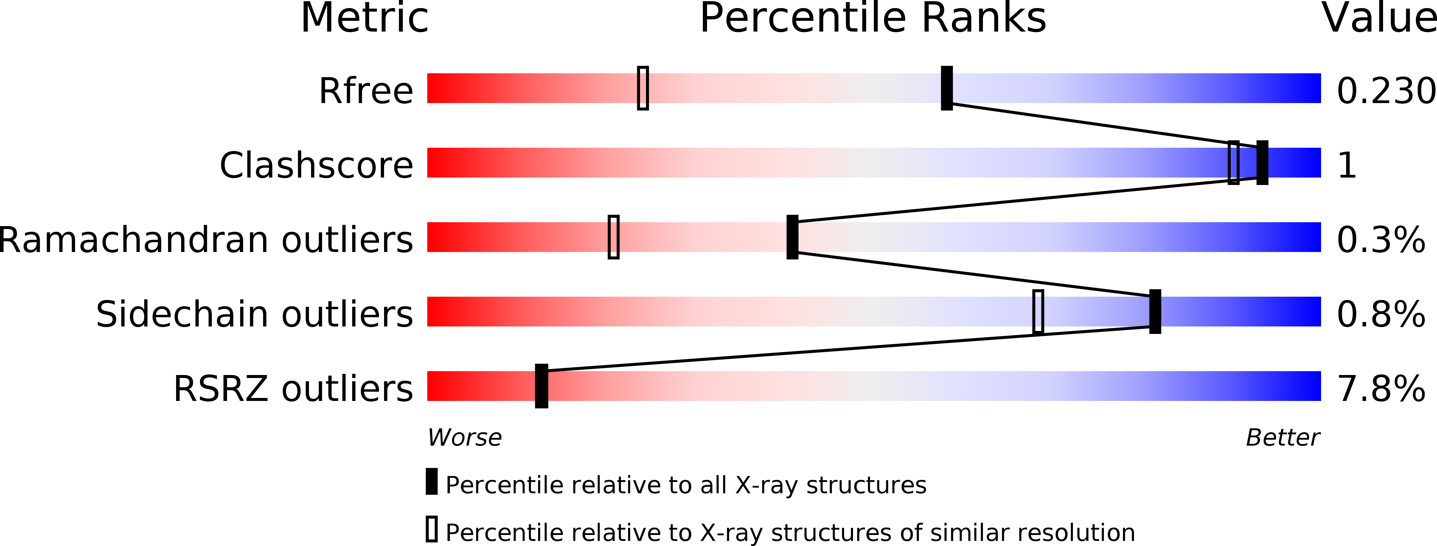
Resolution:
1.58 Å
R-Value Free:
0.21
R-Value Work:
0.20
R-Value Observed:
0.20
Space Group:
H 3
Macromolecular Entities
Polymer Type:polypeptide(L)
Molecule:Biphenyl dioxygenase subunit alpha
Gene (Uniprot):bphA
Chain IDs:A
Chain Length:457
Number of Molecules:1
Biological Source:Comamonas testosteroni
Polymer Type:polypeptide(L)
Molecule:Biphenyl dioxygenase subunit beta
Gene (Uniprot):bphE
Chain IDs:B
Chain Length:186
Number of Molecules:1
Biological Source:Comamonas testosteroni
Primary Citation
Structural Characterization of Pandoraea pnomenusa B-356 Biphenyl Dioxygenase Reveals Features of Potent Polychlorinated Biphenyl-Degrading Enzymes
Plos One 8 e52550 e52550 (2013)
PMID: 23308114 DOI: 10.1371/journal.pone.0052550

Abstact

The oxidative degradation of biphenyl and polychlorinated biphenyls (PCBs) is initiated in Pandoraea pnomenusa B-356 by biphenyl dioxygenase (BPDO(B356)). BPDO(B356), a heterohexameric (αβ)(3) Rieske oxygenase (RO), catalyzes the insertion of dioxygen with stereo- and regioselectivity at the 2,3-carbons of biphenyl, and can transform a broad spectrum of PCB congeners. Here we present the X-ray crystal structures of BPDO(B356) with and without its substrate biphenyl 1.6-Å resolution for both structures. In both cases, the Fe(II) has five ligands in a square pyramidal configuration: H233 Nε2, H239 Nε2, D386 Oδ1 and Oδ2, and a single water molecule. Analysis of the active sites of BPDO(B356) and related ROs revealed structural features that likely contribute to the superior PCB-degrading ability of certain BPDOs. First, the active site cavity readily accommodates biphenyl with minimal conformational rearrangement. Second, M231 was predicted to sterically interfere with binding of some PCBs, and substitution of this residue yielded variants that transform 2,2'-dichlorobiphenyl more effectively. Third, in addition to the volume and shape of the active site, residues at the active site entrance also apparently influence substrate preference. Finally, comparison of the conformation of the active site entrance loop among ROs provides a basis for a structure-based classification consistent with a phylogeny derived from amino acid sequence alignments.

Legend

Protein

Chemical

Disease

Primary Citation of related structures