3GXR image
Deposition Date 2009-04-02
Release Date 2009-10-20
Last Version Date 2023-11-01
Entry Detail
PDB ID:
3GXR
Keywords:
Title:
The crystal structure of g-type lysozyme from Atlantic cod (Gadus morhua L.) in complex with NAG oligomers sheds new light on substrate binding and the catalytic mechanism. Structure with NAG to 1.7
Biological Source:
Source Organism:
Gadus morhua (Taxon ID: 8049)
Host Organism:
Method Details:
Experimental Method:
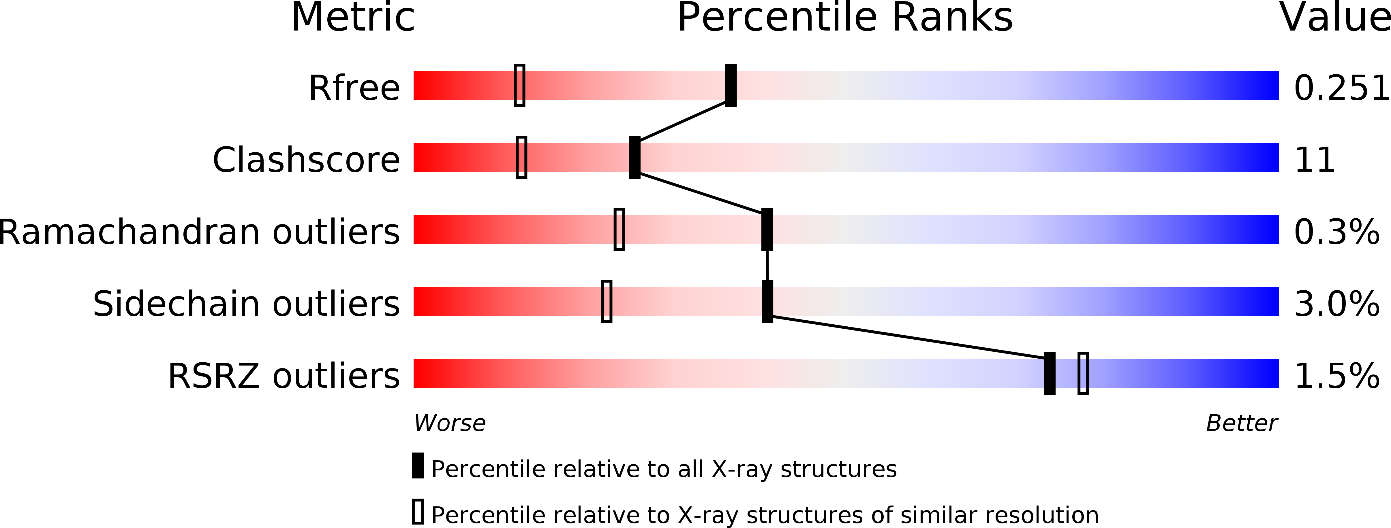
Resolution:
1.70 Å
R-Value Free:
0.25
R-Value Work:
0.20
R-Value Observed:
0.20
Space Group:
C 1 2 1
Macromolecular Entities
Polymer Type:polypeptide(L)
Molecule:Goose-type lysozyme 1
Chain IDs:A, B, C, D
Chain Length:187
Number of Molecules:4
Biological Source:Gadus morhua
Peptide-like Molecules
PRD_900017
Primary Citation
Crystal structures of g-type lysozyme from Atlantic cod shed new light on substrate binding and the catalytic mechanism.
Cell.Mol.Life Sci. 66 2585 2598 (2009)
PMID: 19543850 DOI: 10.1007/s00018-009-0063-x

Abstact

Crystal structures of Atlantic cod lysozyme have been solved with and without ligand bound in the active site to 1.7 and 1.9 A resolution, respectively. The structures reveal the presence of NAG in the substrate binding sites at both sides of the catalytic Glu73, hence allowing the first crystallographic description of the goose-type (g-type) lysozyme E-G binding sites. In addition, two aspartic acid residues suggested to participate in catalysis (Asp101 and Asp90) were mutated to alanine. Muramidase activity data for two single mutants and one double mutant demonstrates that both residues are involved in catalysis, but Asp101 is the more critical of the two. The structures and activity data suggest that a water molecule is the nucleophile completing the catalytic reaction, and the roles of the aspartic acids are to ensure proper positioning of the catalytic water.

Legend

Protein

Chemical

Disease

Primary Citation of related structures