3GXO image
Deposition Date 2009-04-02
Release Date 2010-04-21
Last Version Date 2024-11-20
Entry Detail
PDB ID:
3GXO
Keywords:
Title:
Structure of the Mitomycin 7-O-methyltransferase MmcR with bound Mitomycin A
Biological Source:
Source Organism:
Host Organism:
Method Details:
Experimental Method:
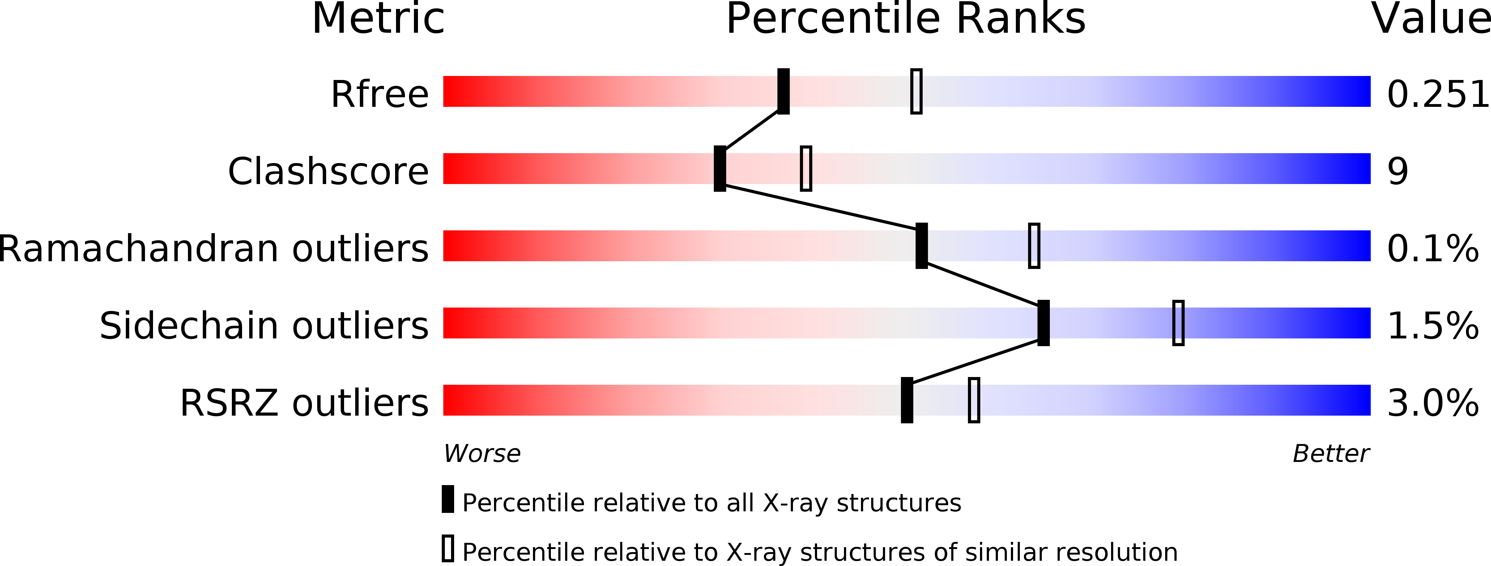
Resolution:
2.30 Å
R-Value Free:
0.25
R-Value Work:
0.20
R-Value Observed:
0.20
Space Group:
P 21 21 21
Macromolecular Entities
Structures with similar UniProt ID
Protein Blast
Polymer Type:polypeptide(L)
Molecule:MmcR
Gene (Uniprot):mmcR
Chain IDs:A, B (auth: D), C, D (auth: B)
Chain Length:369
Number of Molecules:4
Biological Source:Streptomyces lavendulae
Modified Residue
Compound ID Chain ID Parent Comp ID Details 2D Image
MSE A MET SELENOMETHIONINE
Primary Citation
Structural characterization of the mitomycin 7-O-methyltransferase.
Proteins 79 2181 2188 (2011)
PMID: 21538548 DOI: 10.1002/prot.23040

Abstact

Mitomycins are quinone-containing antibiotics, widely used as antitumor drugs in chemotherapy. Mitomycin-7-O-methyltransferase (MmcR), a key tailoring enzyme involved in the biosynthesis of mitomycin in Streptomyces lavendulae, catalyzes the 7-O-methylation of both C9β- and C9α-configured 7-hydroxymitomycins. We have determined the crystal structures of the MmcR-S-adenosylhomocysteine (SAH) binary complex and MmcR-SAH-mitomycin A (MMA) ternary complex at resolutions of 1.9and 2.3 Å, respectively. The study revealed MmcR to adopt a common S-adenosyl-L-methionine-dependent O-methyltransferase fold and the presence of a structurally conserved active site general acid-base pair is consistent with a proton-assisted methyltransfer common to most methyltransferases. Given the importance of C7 alkylation to modulate mitomycin redox potential, this study may also present a template toward the future engineering of catalysts to generate uniquely bioactive mitomycins.

Legend

Protein

Chemical

Disease

Primary Citation of related structures