3GX0 image
Deposition Date 2009-04-01
Release Date 2009-07-07
Last Version Date 2024-02-21
Entry Detail
PDB ID:
3GX0
Keywords:
Title:
Crystal Structure of GSH-dependent Disulfide bond Oxidoreductase
Biological Source:
Source Organism:
Host Organism:
Method Details:
Experimental Method:
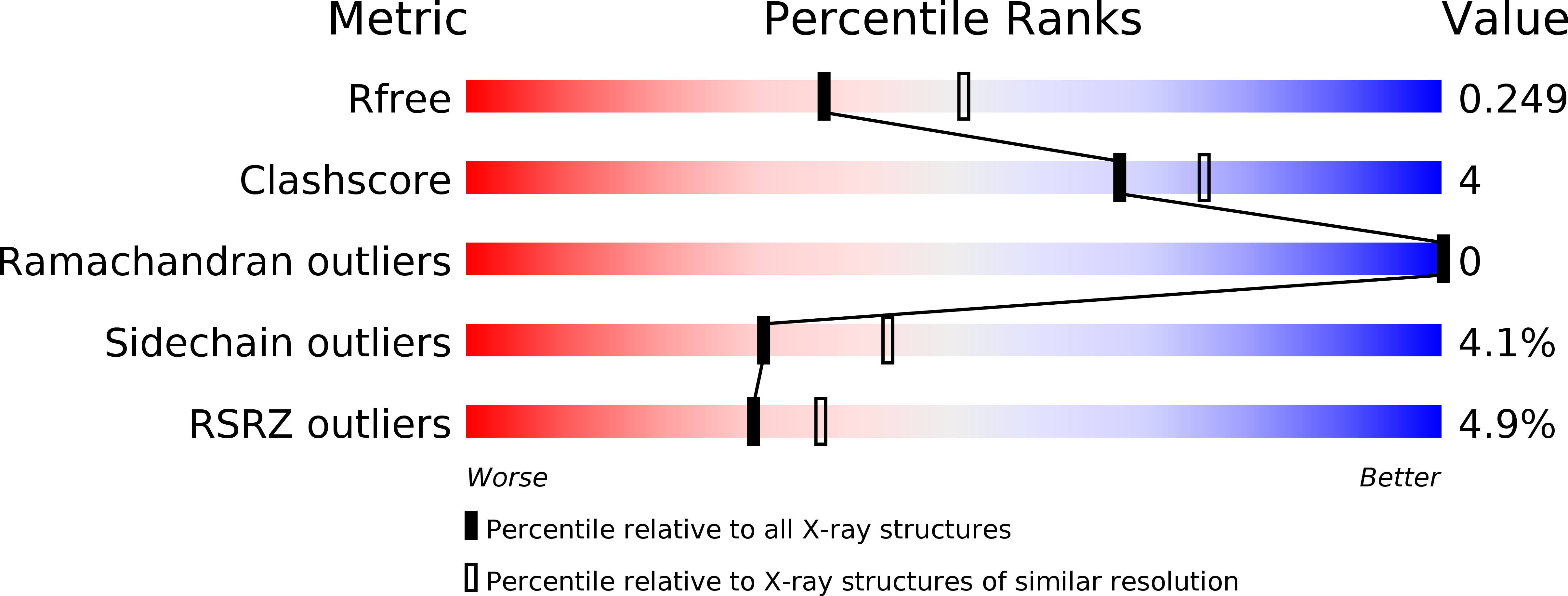
Resolution:
2.30 Å
R-Value Free:
0.24
R-Value Work:
0.19
R-Value Observed:
0.19
Space Group:
P 41 21 2
Macromolecular Entities
Polymer Type:polypeptide(L)
Molecule:GST-like protein yfcG
Gene (Uniprot):yfcG
Chain IDs:A
Chain Length:215
Number of Molecules:1
Biological Source:Escherichia coli
Ligand Molecules
Primary Citation
Analysis of the structure and function of YfcG from Escherichia coli reveals an efficient and unique disulfide bond reductase.
Biochemistry 48 6559 6561 (2009)
PMID: 19537707 DOI: 10.1021/bi9008825

Abstact

YfcG is one of eight glutathione (GSH) transferase homologues encoded in the Escherichia coli genome. The protein exhibits low or no GSH transferase activity toward a panel of electrophilic substrates. In contrast, it has a very robust disulfide-bond reductase activity toward 2-hydroxyethyldisulfide on par with mammalian and bacterial glutaredoxins. The structure of YfcG at 2.3 A-resolution from crystals grown in the presence of GSH reveals a molecule of glutathione disulfide in the active site. The crystallographic results and the lack of functional cysteine residues in the active site of YfcG suggests that the reductase activity is unique in that no sulfhydryl groups in the YfcG protein are covalently involved in the redox chemistry.

Legend

Protein

Chemical

Disease

Primary Citation of related structures