
Deposition Date
2009-04-01
Release Date
2009-04-28
Last Version Date
2023-11-15
Entry Detail
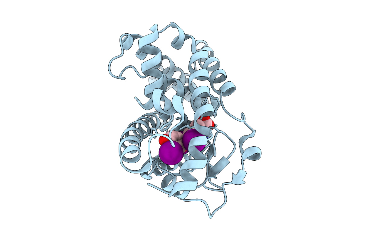
PDB ID:
3GWS
Keywords:
Title:
Crystal Structure of T3-Bound Thyroid Hormone Receptor
Biological Source:
Source Organism(s):
Homo sapiens (Taxon ID: 9606)
Expression System(s):
Method Details:
Experimental Method:
Resolution:
2.20 Å
R-Value Free:
0.23
R-Value Work:
0.19
R-Value Observed:
0.19
Space Group:
P 31 2 1
