3GWI image
Deposition Date 2009-04-01
Release Date 2009-12-15
Last Version Date 2024-03-20
Entry Detail
PDB ID:
3GWI
Keywords:
Title:
Crystal Structure of Mg-ATPase Nucleotide binding domain
Biological Source:
Source Organism:
Host Organism:
Method Details:
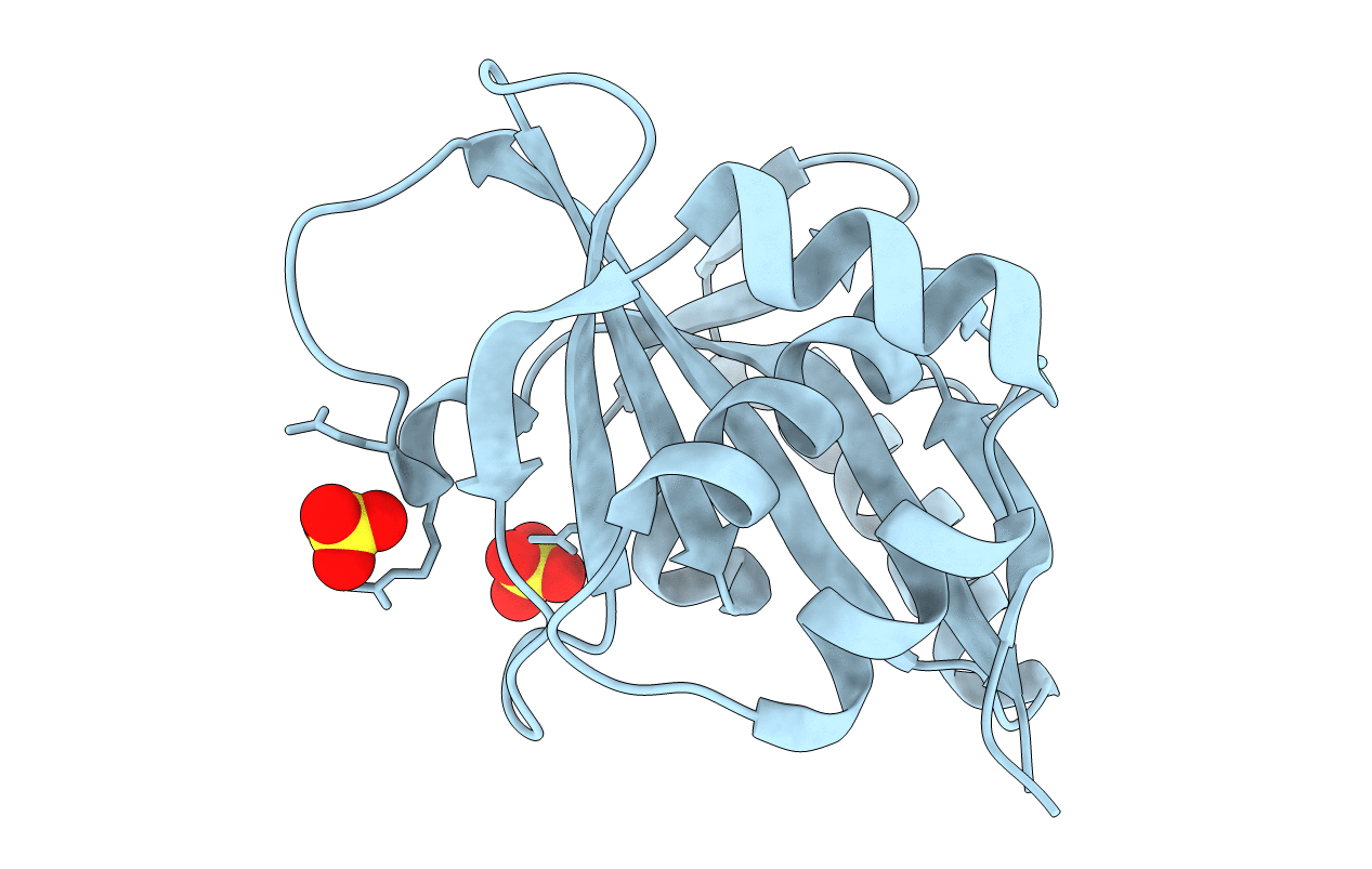
Experimental Method:
Resolution:
1.60 Å
R-Value Free:
0.21
R-Value Work:
0.19
R-Value Observed:
0.19
Space Group:
P 41 21 2
Macromolecular Entities
Structures with similar UniProt ID
Protein Blast
Polymer Type:polypeptide(L)
Molecule:Magnesium-transporting ATPase, P-type 1
Gene (Uniprot):mgtA
Chain IDs:A
Chain Length:170
Number of Molecules:1
Biological Source:Escherichia coli
Ligand Molecules
Primary Citation
The structure of Mg-ATPase nucleotide-binding domain at 1.6 A resolution reveals a unique ATP-binding motif
Acta Crystallogr.,Sect.D 65 1181 1186 (2009)
PMID: 19923713 DOI: 10.1107/S090744490903306X

Abstact

The structure of the nucleotide-binding domain of the Mg-ATPase MgtA from Escherichia coli has been solved and refined to 1.6 A resolution. The structure is made up of a six-stranded beta-sheet and a bundle of three alpha-helices, with the nucleotide-binding site sandwiched in between. The MgtA nucleotide-binding domain is shorter and more compact compared with that of the related Ca-ATPase and lacks one of the beta-strands at the edge of the beta-sheet. The ATP-binding pocket is surrounded by three sequence and structural motifs known from other P-type ATPases and a fourth unique motif that is found only in Mg-ATPases. This motif consists of a short polypeptide stretch running very close to the ATP-binding site, while in Ca-ATPase the binding site is more open, with the corresponding polypeptide segment folded away from the active site.

Legend

Protein

Chemical

Disease

Primary Citation of related structures