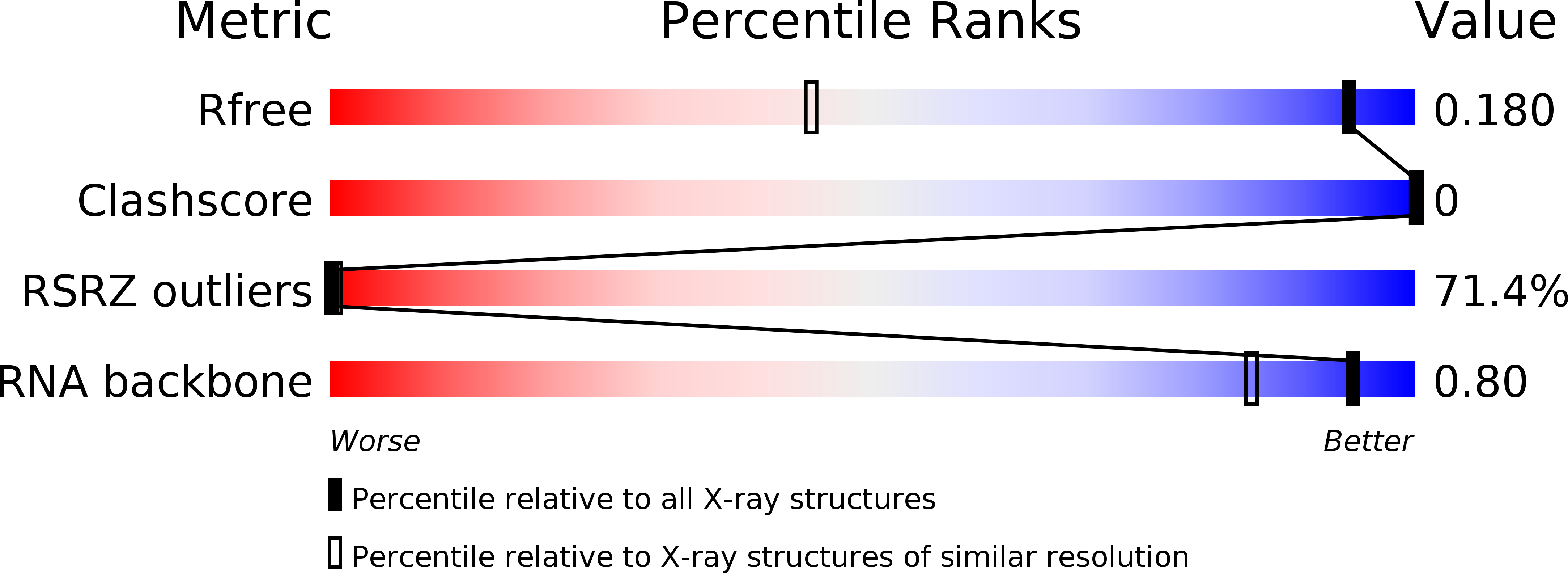
3GVN image
Deposition Date 2009-03-31
Release Date 2009-07-28
Last Version Date 2023-11-01
Entry Detail
PDB ID:
3GVN
Keywords:
Title:
The 1.2 Angstroem crystal structure of an E.coli tRNASer acceptor stem microhelix reveals two magnesium binding sites
Biological Source:
Source Organism:
Method Details:
Experimental Method:
Resolution:
1.20 Å
R-Value Free:
0.20
R-Value Work:
0.19
Space Group:
C 1 2 1
Macromolecular Entities
Polymer Type:polyribonucleotide
Molecule:5'-R(*GP*GP*UP*GP*AP*GP*G)-3'
Chain IDs:A
Chain Length:7
Number of Molecules:1
Biological Source:
Polymer Type:polyribonucleotide
Molecule:5'-R(*CP*CP*UP*CP*AP*CP*C)-3'
Chain IDs:B
Chain Length:7
Number of Molecules:1
Biological Source:
Ligand Molecules
Primary Citation
The 1.2A crystal structure of an E. coli tRNASer)acceptor stem microhelix reveals two magnesium binding sites.
Biochem.Biophys.Res.Commun. 386 368 373 (2009)
PMID: 19527687 DOI: 10.1016/j.bbrc.2009.06.048

Abstact

tRNA identity elements assure the correct aminoacylation of tRNAs by the cognate aminoacyl-tRNA synthetases. tRNA(Ser) belongs to the so-called class II system, in which the identity elements are rather simple and are mostly located in the acceptor stem region, in contrast to 'class I', where tRNA determinants are more complex and are located within different regions of the tRNA. The structure of an Escherichia coli tRNA(Ser) acceptor stem microhelix was solved by high resolution X-ray structure analysis. The RNA crystallizes in the space group C2, with one molecule per asymmetric unit and with the cell constants a=35.79, b=39.13, c=31.37A, and beta=111.1 degrees . A defined hydration pattern of 97 water molecules surrounds the tRNA(Ser) acceptor stem microhelix. Additionally, two magnesium binding sites were detected in the tRNA(Ser) aminoacyl stem.

Legend

Protein

Chemical

Disease

Primary Citation of related structures