Deposition Date
2009-03-30
Release Date
2009-08-25
Last Version Date
2023-09-06
Entry Detail
PDB ID:
3GUI
Keywords:
Title:
T4 lysozyme M102E/L99A mutant with buried charge in apolar cavity--Apo structure
Biological Source:
Source Organism(s):
Enterobacteria phage T4 (Taxon ID: 10665)
Expression System(s):
Method Details:
Experimental Method:
Resolution:
1.45 Å
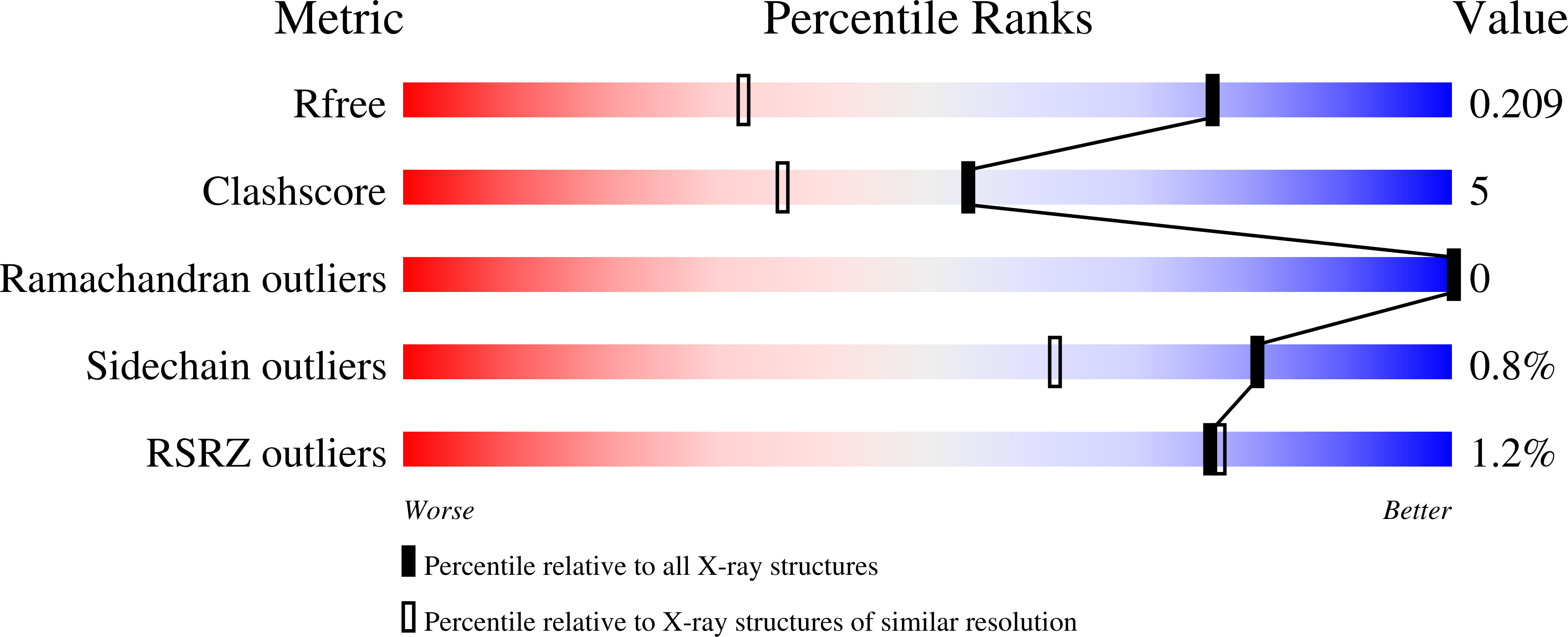
R-Value Free:
0.20
R-Value Work:
0.18
R-Value Observed:
0.18
Space Group:
P 41 21 2